Download
1 / 25
250 likes | 280 Views
Learn about the human heart's anatomy, protective coverings, and blood flow to understand its role as a vital organ. Explore cardiac muscle, the pacemaker, and ECG waves for comprehensive knowledge.

E N D
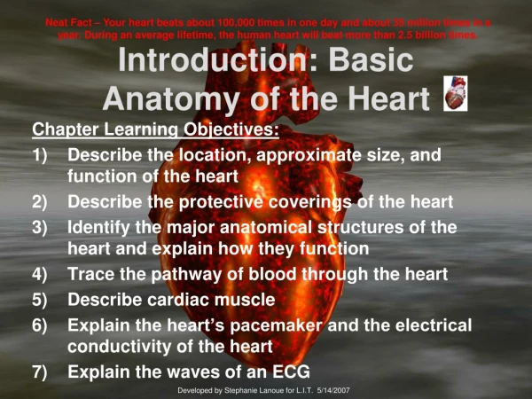
Neat Fact – Your heart beats about 100,000 times in one day and about 35 million times in a year. During an average lifetime, the human heart will beat more than 2.5 billion times. Introduction: Basic Anatomy of the Heart Chapter Learning Objectives: Describe the location, approximate size, and function of the heart Describe the protective coverings of the heart 3) Identify the major anatomical structures of the heart and explain how they function Trace the pathway of blood through the heart Describe cardiac muscle Explain the heart’s pacemaker and the electrical conductivity of the heart Explain the waves of an ECG Developed by Stephanie Lanoue for L.I.T. 5/14/2007
Where is Your Heart Located? Try this: 1)Stand up. 2)Put your hand on your heart (keep it there). Neat Fact: The heart pumps about 1 million barrels of blood during an average lifetime--that's enough to fill more than 3 super tankers.
PREDICT How big is your heart? Neat Fact: On average, your body has about 5 liters of blood continually traveling through it by way of the circulatory system.
How Would You Describe the Heart? What Do You Already Know About It? Class Activity: Appreciating Your Heart as a Pump.
The Heart’s Protective Coverings ▪ Do you know that the heart has 2 protective membranes around it? Heart enclosed in double-walled sac called the ________________. Superficial – fibrous pericardium Deep – serous pericardium *between the 2 layers is some fluid
Remember the number… 4 Next, we are going to learn basic heart anatomy: ▪ 4 chambers ▪ 4 valves ▪ 4 main blood vessels coming into/ exiting the heart
Orientation of the Heart in the Body Right Right Left Left Remember, you are looking at someone else’s chest! (Speaking of someone else’s chest…heart surgery photos)
Photos from Open Heart Surgery Observations – Methodist Hospital, Houston, TX
Basic Heart Anatomy NOTE TO STUDENTS: Label the structures of the heart on your diagram as I explain them. Heart Chambers S. Lanoue NOTE: Atria = plural form of atrium
Heart Anatomy Continued Blood Vessels S. Lanoue
Valves Valves, flaps of connective tissue between the atria and ventricles, allow blood to flow one-way 1 2 3 semilunar valve image (3 cusps are open) mitral valve image ( 2 cusps) S. Lanoue Neat Fact: Heart sounds (“lub, dub”) are caused by the closing of the heart valves. The mitral & tricuspid valves make the 1st sound, and the aortic & pulmonary valves the 2nd.
Valves continued S. Lanoue Chordae tendineae (“heart strings”) attached to ______ and _____ valves. They anchor valve cusps to the papillary muscles (which play a role in valve function). Papillary muscles protrude from the ventricle walls.
12 1 11 10 2 9 3 8 4 7 5 6 REVIEW S. Lanoue
What is a Heart Attack? http://www.healthscout.com/animation/68/13/main.html S. Lanoue
Additional Important Arteries Coronary circulation is blood provided by the right and left coronary arteries. • Left coronary • anterior interventricular artery (also known as the left anterior descending which supplies to the interventricular septum and anterior walls of both ventricles) • circumflex artery (supplies left atrium and posterior walls of left ventricle). • Right coronary • marginal artery (supplies myocardium of the lateral right side) • posterior interventricular artery
Cardiac Veins • After passing through capillary beds of the myocardium, the blood flows into the coronary sinus (which empties blood into right atrium). Has 3 tributaries: • ______ cardiac vein • ______ cardiac vein • _______ cardiac vein Also, several anterior cardiac veins.
Partner Activity: Blood Flow Through the Heart Instructions: Working with a partner, can you describe the flow of blood through the heart? Start with the right atrium. List all the major structures along the way (chambers, valves, and vessels). Refer to the diagram on page 602. Example: Rt. atrium > ? valve> ? (chamber) > ? valve up the pulmonary trunk which divides into the > ? arteries (which send the deoxygenated blood into the ? for O2.) From the lungs back to the heart along the ? Veins > ? (chamber) through the ? Valve >? (chamber) > ? Valve > Aorta > Body > Venae cavae.
Cardiac Muscle Cardiac muscle – _______, short (fat/ _______) cells that are interconnected for quicker communication & conduction of electrical current. Video footage: http://video.aol.com/video-detail/beating-human-heart-cells-from-embryonic-stem-cells/4230805508
Conduction System Know Sequence of Excitation: 1. Sinoatrial node (SAN) or __________ > 2. _______ventricular node (AVN) > 3. Atrioventricular bundle (bundle of ___) > 4. Right and Left bundle branches > 5. Purkinje fibers (directly supply the papillary muscles) See animation: http://www.jdaross.cwc.net/cardiac_cycle.htm
Electrocardiogram (ECG) http://www.cardioconnection.org/frameWork.aspx?cnt=cad/diagnosis/ecg# 3 Distinguishable Waves: ___ – initiation of the heartbeat in the atria ___ – movement of the electrical current through the ventricles __ – recovery phase; electrical current spreads back over the ventricles in the opposite direction
Electrocardiogram (ECG) Findings Healthy heart – _____, ______, and timing of waves tend to be _____________ Unhealthy heart – waves are ______ Examples: an enlarged R wave hints of enlarged ventricles or a prolonged Q-T interval may reveal ventricular arrhythmias
Normal and Abnormal ECG Readings • _______– steady sinus rhythm • Non-functional _____ • Heart _____ – damage to the AVN (interferes with the ventricles receiving pacing impulses) • __________ fibrillation (acute heart attack or electrical shock)
Normal and Abnormal ECG Readings continued Arrhythmias – ________ heartbeats caused by uncoordinated atrial and ventricular contractions Example: atrial flutter http://www.youtube.com/watch?v=3q4TJUmMztY ___________ – rapid and irregular contractions; SAN looses control of setting the pace; fatal condition
Other Heart Abnormalities Heart ________ – blood back flowing through a heart valve ___________ –abnormally fast heart rate __________ – heart rate slower than 60 beats per minute Congestive heart failure – progressively worsening condition in which the heart can no longer pump blood efficiently (heart weakens and enlarges)