به نام خدا
290 likes | 430 Views
به نام خدا. Atelectasis & adhesive otitis. Kayvan Aghazadeh M.D Assistant prof. of otolaryngology Tehran university of medical siences Amir Alam hospital . Middle ear atelectasis is thought to result mainly from long-standing eustachian tube dysfunction.

به نام خدا
E N D
Presentation Transcript
Atelectasis & adhesive otitis • KayvanAghazadeh M.D • Assistant prof. of otolaryngology • Tehran university of medical siences • Amir Alam hospital
Middle ear atelectasis is thought to result mainly from long-standing eustachian tube dysfunction. • One of the main functions of the eustachian tube is ventilation of the middle ear and mastoid
Opening of the eustachian tube allows exchanging of gases and equalization between the environment and middle ear. • The middle ear gases also are exchanged with the middle ear mucosa.
Bilateral diffusion between the middle ear cavity and the blood may be an important factor in middle ear atelectasis because: • the gas composition of the middle ear basically resembles that of venous blood.
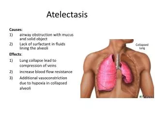
If the atelectasis develops, the tympanic membrane becomes retracted onto the promontory and the ossicles of the middle ear.
In atelectatic ears, the middle ear space is partially or completely obliterated, but the tympanic membrane is not adherent to the medial wall of the middle ear, • and the mucosal lining of the middle ear is intact
In contrast, adhesive otitis media exists when the middle ear space is totally obliterated, • and the tympanic membrane is adherent to the ossicles and promontory; • mucosal surfaces are not present.
Retraction of the tympanic membrane may lead to erosion of the long process of the incus and the stapes suprastructure
Not all patients with chronic OME develop atelectasis; in most patients with OME, retraction of the tympanic membrane is limited. • In patients with bilateral OME, 1.5% of untreated ears and 2% of ears treated with tubes developed severe atelectasis.
It may be that repeated bouts of AOM lead to weakening and thinning of the membrane, which allows atelectasis
Sad and Bercoshowed destruction of the collagen-containing fibrous layer of the tympanic membrane in some ears with recurrent infection. • Collagen destruction within the tympanic membrane may lead to another complication of OME—tympanosclerosis
. Sad and Bercoand Tos and Poulsendescribed four stages of tympanic membrane retraction: • stage I, retracted tympanic membrane; • stage II, retraction with contact onto the incus; • stage III, middle ear atelectasis; and • stage IV, adhesive otitis media
Middle ear atelectasis may be reversible with ventilating tubes. • Sad showed that ventilating tubes improved the state of atelectatic ears.
Graham and Knightreported three cases in which atelectatic tympanic membranes were restored to their normal position • by administration of nitrous oxide during anesthesia and insertion of a ventilating tube.
Atelectasis and adhesive otitis media usually coexist with OME, • although OME may resolve in these ears, allowing aeration of the attic and mastoid, but leaving a collapsed middle ear.
In extreme cases, when hearing loss or ossicular erosion occurs, • a myringoplasty for the reinforcement of atelectatic tympanic membrane may be indicated.
Cholesteatomas may originate from deep retraction pockets in which desquamated keratin debris would not be cleared into the ear canal
These retraction pockets may occur in the pars tensa or pars flaccida of atelectatic ears, • and should be considered precursors to cholesteatomas
Nonpneumatized mastoids may have a limited ability to buffer pressure changes and • can manifest as an atelectasis, a retraction pocket, or a cholesteatoma