Download

1 / 37
520 likes | 2.21k Views
Atelectasis . Sam Wasmann. Atelectasis:. Collapse or loss of lung volume May involve entire lung, a lobe, a segment, or be subsegmental There are 5 mechanisms of atelectasis: 1) (Post) obstructive 2-5) Non-obstructive – typically due to loss of contact between parietal and visceral pleura.

E N D
Atelectasis Sam Wasmann
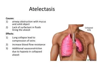
Atelectasis: • Collapse or loss of lung volume • May involve entire lung, a lobe, a segment, or be subsegmental • There are 5 mechanisms of atelectasis: • 1) (Post) obstructive • 2-5) Non-obstructive – typically due to loss of contact between parietal and visceral pleura. • This presentation will cover the 5 mechanisms of atelectasis, as well as radiographic findings of collapse in all 5 lung lobes.
Atelectasis Does Not Cause Fever • In a 1995 study of 100 postoperative cardiac surgery patients receiving daily portable chest x-rays,“no association could be found between fever and amount of atelectasis. This contradicts common textbook dogma but agrees with previous human study and animal experiments.” Engoren, Milo. “Lack of Association Between Atelectasis and Fever.” Chest. Volume 107(1) January 1995 pp 81-84 • A 1988 study of 270 patients after elective intra-abdominal surgery found that “neither the presence nor the absence of fever can assure clinicians of the presence or absence of a postoperative pathologic pulmonary complication such as atelectasis.” Roberts J, Barnes W, Pennock M, Browne GD. “Diagnostic Accuracy of Fever as a Measure of Postoperative Pulmonary Complications.” Heart Lung. 1988 Mar;17(2):166-70
CXR Findings in Atelectasis • Direct: • Displacement of fissures • Increased opacification of airless lobe • Crowded air bronchograms (non-obstructive only) or vessels • Indirect: • Displacement of hilar structures • Ipsilateral cardiomediastinal shift • Narrowing of ipsilateral intercostal spaces • Obscured structures adjacent to atelectasis • Elevation of ipsilateral diaphragmatic leaflet • Hyperexpansion/hyperlucency of remaining aerated lung
Typical findings of atelectasis in this patient include: 1) Hazy opacity in left upper lung (direct sign) 2) Left tracheal shift (indirect sign) 3) Loss of left cardiac silhouette (indirect sign)
Right Lobar Anatomy Approximate position of right upper, middle and lower lobes on chest x-ray.
Right Lobar Anatomy Lateral View
Left Lobar Anatomy Approximate position of left upper and lower lobes on chest x-ray.
Left Lobar Anatomy Lateral View
Right Upper Lobe Atelectasis Findings include: • Elevation of right hilum and minor fissure • Collapsed lobe shifts cephalad and medially • If due to a central mass, the minor fissure retracts cranially with a lateral upward convexity and a medial caudal convexity (S-sign of Golden). This suggests neoplastic etiology.
Right upper lobe atelectasis: The atelectatic RUL forms a triangular opacity (arrow). The elevated minor fissure is retracted cranially (see image below) and forms a reverse S shape (S-sign of Golden) as it curves around the hilar mass (M).
Right Middle Lobe Atelectasis • Right middle lobe is only 10% of total lung volume. • Greater tendency to collapse than other lobes. • Radiographic findings can be subtle: • Small triangular opacity pointing laterally • Obscured right heart border • Lateral view: obliquely oriented triangular opacity with apex pointed toward hilum.
Right middle lobe atelectasis: There is a small triangular opacity pointing laterally, right cardiac border is partially obscured, and slightly lower lung volume in right compared to left.
Lateral view: The arrows point to the major and minor fissures which are parallel to each other. The atelectatic middle lobe is the opacity between the fissures. Notice that it projects over the cardiac silhouette.
Right Lower Lobe Atelectasis • When atelectatic, right lower lobe retracts posteromedially and inferiorly. • Major fissure is shifted downward and becomes visible • As RLL collapses, it forms a triangular opacity which obscures the left lobe pulmonary artery, and eventually forms a right paraspinal mass that projects behind the right atrium. • On lateral view, posterior 1/3 of right diaphragm is obscured by collapsed RLL. Diaphragm may not be obscured on frontal view because hyperexpanded middle lobe abuts it.
RLL Atelectasis: Triangular opacity in right lower hemithorax. The lateral border is the major fissure (not normally seen on frontal view). Right hilum is displaced caudally and partially obscured. The hyperexpanded RML outlines the cardiac border and right hemidiaphragm.
Left Upper Lobe Atelectasis • Faint, hazy opacity in left upper hemithorax • 50% of patients have complete major fissure • Main pulmonary trunk and upper contour of left pulmonary artery are obliterated • Left hilar structures and left lower lobe are retracted caudally (look for superior segment vessels from the lower lobe occupying the apex, mimicking an aerated upper lobe) • 50% have an incomplete major fissure • Tongue of aerated lower lobe is pulled forward by atelectatic lobe, between the atelectasis and the aortic arch, forming a crescent-shaped lucency (Luftsichel sign) • Diaphragm typically elevated
Left upper lobe atelectasis: Opacity contiguous to the aortic arch. The mediastinum is shifted toward the left hemithorax, which is small in comparison to the right.The main pulmonary trunk and the left pulmonary artery are obliterated.
Left upper lobe atelectasis in patient with incomplete major fissure: There is an ill-defined opacity in the left half of the left upper thorax. The trachea is deviated left and the left hilum is retracted superiorly. Vascular branches to the left lower lobe superior segment form an array of linear and tubular opacities. The arrow shows a vertical lucency separating the aortic arch from the vertical margin of the collapsed lobe (Luftsichel).
Left Lower Lobe Atelectasis • Common after cardiac surgery • Radiographic findings include: • Increased retrocardiac opacity • Obscuring of the left lower lobe vessels and left hemidiaphragm • Caudal displacement of left hilum • Levorotation of cardiac silhouette with flattening of cardiac waist • Mediastinal shift can cause partial obliteration of aortic arch
LLL Atelectasis: Notice the wedge shaped opacity behind the cardiac silhouette. The border is formed by the major fissure (arrow). The left hilum is partially obscured and displaced caudally. The left upper lobe is hyperexpanded accounting for the increased lucency in the left hemithorax.
Complete Atelectasis of Entire Lung • Total collapse of a lung • Complete opacification of an entire hemithorax • Ipsilateral cardiomediastinal shift (in massive pleural effusion, would shift to contralateral side). • Cardiac silhouette, one hemidiaphragm, and one hilum are obscured in lateral projection.
Complete left lung atelectasis: There is mediastinal displacement, opacification, and loss of volume in the left hemithorax. The cardiac silhouette (which is shifted left) is obscured, as are the left hilum and left hemidiaphragm.
Obstructive (Resorptive) Atelectasis • Most common type • Results from blockage of airway • mucous plugging, foreign body, neoplasm, or inflammatory debris • Air distal to obstruction is resorbed from nonventilated alveoli • Findings include loss of lung volume without presence of air bronchograms
Post-obstructiveatelectasis of RLL: The major fissure is visible as it has rotated into view. There are no air bronchograms seen within the atelectatic region of lung. The patient is intubated. The obstruction is likely due to mucous plugging.
Non-obstructive Atelectasis 1) Passive 2) Compressive 3) Cicatrization 4) Adhesive In these forms of atelectasis secretions are able drain up the bronchial tree. Because there is no obstruction, bronchoscopy is not therapeutic.
Passive (Relaxation) Atelectasis • 2nd most common form of atelectasis • Contact between parietal and visceral pleura is lost due to pleural effusion or pneumothorax. • Leads to generalized collapse.
Passive atelectasis: Notice the crowded air bronchograms (arrows) in the setting of a left pleural effusion. Air bronchograms are not present in post-obstructive atelectasis.
Compressive Atelectasis • Due to external compression of lung • May be caused by loculated collection of pleural fluid or by masses in chest wall, pleura, or parenchyma. • Similar to relaxation atelectasis but collapse is local rather than generalized.
Compressive atelectasis: Chest x-ray showing a giant bulla occupying more than two thirds of the right hemithorax and compressing the underlying lung upward and toward the mediastinum. Crowded air bronchograms can be seen (arrows).
Adhesive Atelectasis • Caused by adherence of the alveolar wall surfaces in the setting of surfactant deficiency (e.g., hyaline membrane disease) • Surfactant has phospholipid dipalmitoyl phosphatidylcholine, which prevents lung collapse by reducing the surface tension of the alveoli • Lack of surfactant or inactive surfactant cause alveolar instability and collapse
Adhesive atelectasis in infant with hyaline membrane disease: CXR reveals bilateral ground-glass appearance of the lungs (atelectasis) and air bronchograms standing (red arrow) out against the collapsed parenchyma.
Cicatrization Atelectasis • Secondary to fibrosis (scarring) of lung parenchyma with subseqent lack of expansion • Etiologies include granulomatous disease (often occurs in sarcoid, fungal, and chronic TB), necrotizing pneumonia, and radiation.
Cicatrization atelectasis: Lung destruction in patient with chronic pulmonary tuberculosis.
References: 1) Sharma, Sat. “Atelectasis.” e-medicine, 2004. http://www.emedicine.com/med/topic180.htm#section~pictures 2) Brad H. Thompson, M.D., William J. Lee, B.S., Jeffrey R. Galvin, M.D. and Jeffrey S. Wilson, M.D “Lobar Anatomy” ElectricLungAnatomy www.vh.org/adult/provider/radiology/LungAnatomy/LobarAnat/LobarAnat.html 3) Daffner, RH. Clinical Radiology – The Essentials. Williams and Wilkins, 1993, pp 80-85. 4) Engoren, Milo. “Lack of Association Between Atelectasis and Fever.” Chest. Volume 107(1) January 1995 pp 81-84 5) Roberts J, Barnes W, Pennock M, Browne GD. “Diagnostic Accuracy of Fever as a Measure of Postoperative Pulmonary Complications.” Heart Lung. 1988 Mar;17(2):166-70 6) Stark, Paul. “Atelectasis: Types and Pathogenesis.” UpToDate, 2004. 7) Stark, Paul. “Radiologic Patterns of Lobar Atelectasis.” UpToDate, 2004. 8) Weed HG, Baddour LM. “Postoperative Fever.” UpToDate, 2004. 9) Federico Venuta and Tiziano de Giacomo. “Giant Bullous Emphysema.” CTSNET Experts' Techniques, General Thoracic Experts' Techniques. http://www.ctsnet.org/doc/6761