Enhanced Canine Disc Diagnosis: Single-Fiber Reflectance Spectroscopy
Investigating the accuracy of single-fiber reflectance spectroscopy versus radiography and CT for identifying degenerated canine intervertebral discs. Detailed background, motivations, imaging techniques, results, and surgical treatments explored. Source links provided for comprehensive understanding.

Enhanced Canine Disc Diagnosis: Single-Fiber Reflectance Spectroscopy
E N D
Presentation Transcript
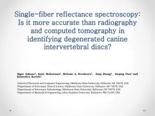
Single-fiber reflectance spectroscopy: Is it more accurate than radiography and computed tomography in identifying degenerated canine intervertebral discs? Nigar Sultana1, Kelci McKeirnan2, Melanie A. Breshears3, Anqi Zhang4, Daqing Piao1 and Kenneth E. Bartels2 1School of Electrical and Computer Engineering, Oklahoma State University, Stillwater, OK 74078, USA 2Depertment of Veterinary Clinical Science, Oklahoma State University, Stillwater, OK 74078, USA 3Depertment of Veterinary Pathobiology, Oklahoma State University, Stillwater, OK 74078, USA 4Depertment of Biomedical Engineering, Johns Hopkins University, Baltimore, MD 21205, USA
OVERVIEW • Background • Motivation and Objective • System configuration • Imaging protocols • Results • Summery
Background • Intervertebral discs (IVD), located between the vertebrae in the spine and act as “shock absorbing cushions” which allows slight movement of vertebrae • Intervertebral discs are comprised of two layers • Annulus fibrosus ---Tough outer fibrous lamellar layer • Nucleus pulposus --- Gelatinous inner core Source: http://www.spineuniverse.com/conditions/herniated-disc
Background • Intervertebral disc herniation is a common disease that affects the vertebra • The chondrodystrophoic dogs (dachshund, Pekingese, etc.) mainly seem to have disc degeneration along the entire vertebral column while it is even common in humans also • Loss of nucleus fluid and deposition of a calcified mineral component within the nucleus makes the disc degenerated • Degenerated disc is less resilient to normal wear and tear • Degeneration can cause severe pain, nerve compression and paralysis Extrusion of degenerated disc material through the dorsal annulus Source: http://www.mayfieldclinic.com/PEHLDisc.htm#.Ujy34sakquM
Background Treatment • Surgical disc fenestration--- most widely used treatment for intervertebral disc disease • It’s a highly invasive procedure • Very complex • Recurrence rate as high as 19.2 % as reported by several large scale studies of a mixed population of dogs Source: http://cal.vet.upenn.edu/projects/saortho/chapter_62/62mast.htm
Background • Percutaneous Laser disc Ablation (PLDA)--- minimal invasive procedure for intervention of intervertebral disc disease • This laser surgical treatment photothermally ablates the disc material and fibrotic scar tissue forms which presumably prevents the disc from herniating in the future • PLDA is performed by passing an optical fiber through a needle placed percutaneously in the center of a disc to introduce laser energy, most frequently from a 2100 nm holmium:YAG (Ho:YAG) laser • PLDA shown to have minimum recurrence rate as low as 3.4 %
Motivation • In current practice, PLDA is performed in thoracolumbar IVD with same laser fluence applied to each disc, regardless of its mineral content • Knowledge of individual disc mineral composition would allow treatment of degenerated discs that otherwise may have been missed • Mineralized volume quantization of individual disc would permit laser energy dosage adjustments for specific degenerated disc
Motivation (Imaging) • Current standard for diagnostic imaging for canine IVD degeneration is X-ray radiography, which usually shows as opacification of the disc space • Radiography is also used to guide the needle positioning into the center of each treated disc for PLDA • However, radiography is not adequately sensitive in identifying the degenerated discs, nor is it reliable for characterizing the degree of disc degeneration • When compared with histopathology, radiography has a sensitivity of 30 % and specificity of 100 % in diagnosing disc degeneration Source: http://www.vetsurgerycentral.com/myelogram.htm
Motivation(imaging) • X-ray computed tomography (CT) and magnetic resonance imaging (MRI) usually able to pinpoint the precise location of degenerated disc • However, it is not also highly sensitive in characterizing the degree of disc degeneration and sometimes provide misleading results in identifying the degenerated discs • When compared with histopathology, computed tomography (CT)has a sensitivity of 75% and specificity of 60 % in diagnosing disc degeneration • Moreover, due to the cost and inconvenience it is challenging to standardize PLDA surgery under CT or MRI image guidance Source: http://www.mayfieldclinic.com/PE-HLDisc.htm#.Ujy7jsakquM http://canadawestvets.com/disc-disease
Motivation (Optical) • An imaging/sensing technology that provides a more accurate pre-operative in-situ assessment of the disc mineralization, • and potentially rapid post-operative feedback, could optimize the outcome of the PLDA procedure • Single-fiber reflectance spectroscopy---- • Sense the changes of tissue compositions in the disc • A diagnosis modality that could be integrated to PLDA procedure http://www.dfwvetsurgeons.com/downloads/Newsflash_rdvm.pdf
Hypothesis (scattering) • The hypotheses • The mineral deposit within degenerated IVD increases scattering • IVD that contains mineral works as an avascular medium • The more degenerated disc is, the more scattering the medium should become
Objective • The objective of this study is to revaluate whether the needle-based single-fiber reflectance spectroscopy could have higher accuracy in identifying degenerated intervertebral discs compared to X-ray radiography and computed tomography (CT) • If needle-based single-fiber reflectance spectroscopy could provide a positive correlation with histopathology results • If SfRS could have a higher sensitivity in grading degree of disc degeneration
System configuration The experimental setup includes: • Halogen -Deuterium Source • Spectrometer • Bifurcated fiber bundle • 320μm single fiber with 15° angle polished tip • Computer • 20 gauge needle Computer Light Source Spectrometer Bifurcated fiber bundle • single fiber 150angle polished fiber in needle 15° Schematic of Single fiber reflectance spectroscopy setup
Analytical Representation ) Raw Spectrum Normalized Spectrum
Imaging protocol Acquisition time: 2 second/measurement -Step1: Measurements -Step2: PLDA procedure -Step3: Measurements extending ~1mm Reflectance spectroscopy system
Results (Dog#1 Radiography and CT) L2-3, slight nuclear mineralization L4-5, no nuclear mineralization No mineral present on radiographs
Results (Dog#2 Radiography and CT) T10-11 mineralized T12-13 mineralized T11-12 mineralized L1-2 mineralized L2-3 mineralized L3-4 mineralized L4-5 mineralized L5-6 mineralized T8-9 T12-12 L2-3 L3-4 L4-5 L7-S1 are mineralized in radiograph
Results (Dog#3 Radiography and CT) T10-11 mineralized T11-12 mineralized T12-13 mineralized T13-L1 mineralized L2-3 mineralized L3-4 mineralized L4-5 mineralized L1-2 mineralized T10-11 mineralized
Results (Radiography and CT) • Radiographic and CT imaging were performed on all thoracic and lumbar intervertebral discs of the two cadaveric dogs • The needles for Single fiber reflectance spectroscopy sensing and subsequently for PLDA were placed successfully in “ Dog 1” from T8– 9 to L5– 6 and in “ Dog 2 and Dog 3” from T9– 10 to L5– 6. Therefore only the radiography and CT images of those discs being evaluated by Single fiber reflectance spectroscopy measurement are displayed
Results • In histopathology result, 20 out of 28 discs were determined to have different level of mineral deposition (5/10 form dog1, 4/9 from dog2 and 9/9 from dog3) • Out of 20 degenerated discs • Radiography identified 6 degenerated discs • CT was successfully identified 15 degenerated discs and • SFRS identified 19 degenerated discs correctly R19 identified by SfRS
Results • Normalization is applied to all 28 discs with respect to air and water from entire optical range of 530-1000nm • Blue dashed line represents normalized spectra of normal disc (T12-13-Dog1, L3-4-Dog1 and L1-2 Dog1) • Red dotted line represents normalized spectra ofdegenerated disc (T12-13-Dog3, T13-L1-Dog3, L1-2-Dog-1, T11-12-Dog1) identified by CT and histopathology
Mineralized Normal Pathology (% of mineral volume) SfRS (avg normalized intensity of 530-1000nm) Computed Tomography (CT) Radiography T8-9 T10-11 T11-12 T12-13 T13-L1 L1-2 L2-3 L3-4 L4-5 L5-6
Results • The comparison of SfRS normalized intensity with radiography and CT based on histopathology result for discs T8-9, T10-11, T11-12, T12-13, T13-L1, L1-2, L2-3, L3-4, L4-5 and L5-6 are shown here for three cadaveric dogs • The red triangle represents degenerated disc and blue circle is the representation of normal disc (not having any mineral) • For SfRS, the normalized intensity with respect to air and water is presented on an average of optical range of 530-1000nm • SfRS result showed a good correlation with histopathology result of different percentage of mineral content by different normalized intensity level
Results T8-9 T10-11 T11-12 T12-13 T13-L1 L1-2 L2-3 L3-4 L4-5 L5-6 • Receiver operating characteristic (ROC) curvefor average normalized spectral intensity of SfRSfrom 530-1000nm is generated by varying threshold level to determine SfRS classification in identifying degenerated disc • Threshold is placed where SfRS could identified 19 out of 20 degenerated discs
Results • ROC curve is the graphical presentation of sensitivity and specificity to determine system classification where, • The dotted line represents a random guess of 50% accuracy • The solid line shows the sensitivity and specificity when SfRS identified 19 out of 20 degenerated disc by threshold placement • Area under ROC curve of SfRSdetermined to be 0.841
Results • SfRS normalized intesity averaged from 530 to 1000nm for all 28 discs are plotted against the percentage amout of mineralized volume determined by histopathology • Up to 50% of mineralized volume, the correlation coefficient, R obtained as 0.8732 and coefficient of determination, determined to be 0.7625 Homogenous Avascular
Summery • In identifying and grading canine intervertebral disc degeneration, Single fiber Reflectance Spectroscopy is performed • A needle-based single-fiber reflectance spectroscopy system that is compatible with PLDA procedure is constructed • SfRS measurements indicate increase of light scattering intensity across the entire 530 –100 nm spectral range in discs with mineralization • In grading disc mineral composition, SfRS showed a higher sensitivity and specificity than both X-ray radiography and computed tomography (CT)
Summery • In identifying degenerated disc by varying threshold level, SfRS showed a good classification with having area under ROC curve of 0.841 • At high percentage of mineral volume (above 50%) confirmed by histopatology imaages, due to higher number of scattering particle, deposited mineral acts as a homogeneous medium and only accounts for fresnel reflection while applying SfRS, thus SfRS could not provide as good correlation as for less than 50%of mineral volume
Acknowledgement • Endowment to Bartels KE, Kerr Foundation, Oklahoma city, OK • Oklahoma Center for the Advancement of Science and Technology (OCAST) HR 11-043