Download
1 / 38
380 likes | 445 Views
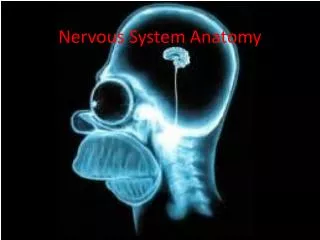
Explore the anatomy of the central nervous system, major divisions, and distinct parts like the cerebral cortex, diencephalon, brain stem, and cerebellum. Learn about grey vs. white matter, sulci, gyri, and sensory receptors. Understand sensory, motor divisions, and their functions. Written by Jeremy J. McLemore, a Pharm.D. Candidate at Howard University College of Pharmacy.

E N D
Anatomy of the Central Nervous System Pharmacy Biomedical Preview July 7, 2015 Jeremy J. McLemore Howard University College of Pharmacy Pharm D Candidate, Class of 2017 APhA | SNPhA | ASHP | ΦΛΣ | KΨ
Notable Differences Size Sulci Gyri Orientation
Anatomical Orientations Fundamental Neuroscience for Basic and Clinical Applications, D.E. Haines (editor), 3rd edition, 2006, Churchill Livingstone Elsevier Publishing, Philadelphia, PA.
Terminology • Ganglia vs nucleus • Tract vs nerve • White matter vs grey matter • Medial vs lateral • Ipsilateral vs contralateral • Planes of section • Coronal • Sagittal • Horizontal (axial) • Tangential
Central vs Peripheral Nervous System Somatic Nervous System Autonomic Nervous System Fundamental Neuroscience for Basic and Clinical Applications, D.E. Haines (editor), 3rd edition, 2006, Churchill Livingstone Elsevier Publishing, Philadelphia, PA. http://www.warriorpages.com/anatomyqigong/anatomy/nervous.html
Major divisions of the CNS • Cerebral Cortex • Diencephalon • Brain Stem • Cerebellum Fundamental Neuroscience for Basic and Clinical Applications, D.E. Haines (editor), 3rd edition, 2006, Churchill Livingstone Elsevier Publishing, Philadelphia, PA.
Cerebral Cortex • Composed of “grey” and “white” matter • Organ of information processing • Divided into hemispheres by one longitudinal fissure • Also divided into 4 distinct lobes
Cerebral cortex is convoluted, creating sulci (grooves) and gyri (bumps) • These foldings are • fairly well preserved across individuals • functionally segregated (Brodmann’s areas) • used as landmarks to partition the cerebral cortex into lobes Lateral Surface Fundamental Neuroscience for Basic and Clinical Applications, D.E. Haines (editor), 3rd edition, 2006, Churchill Livingstone Elsevier Publishing, Philadelphia, PA.
Diencephalon • Located between the cerebral cortex and brain stem • Consists of 3 distinct parts
Thalamus • Sensory relay center between brain stem and cortex • Consciousness, sleep, alertness
Epithalamus • Houses the pineal gland which secretes melatonin • Thought to control emotion and house certain motor pathways
Hypothalamus • Interacts with pituitary gland to regulate metabolic and endocrine function
Brain Stem • Controls vital functioning • Midbrain • Motor control, vision, alertness, temperature • Pons • Motor coordination relay center • Sleep, swallowing, taste, facial movement • Medulla • Breathing, heart rate, vomiting, blood pressure
Cerebellum • Coordinates sensory input with muscular responses (does not initiate movement) • Decerebrate Cat
Peripheral Nervous System • Components outside of the central nervous system • Sensory receptors, ganglia, motor endings, peripheral nerves • Bridge from our CNS to the external environment • Point of communication for PNS?
Sensory Division • Structures specialized for stimulus response • DepolarizationSignaltrasmission • Processing occurs where? • Efferent vs Afferent fibers
5 Types of Sensory Receptors • Chemoreceptor-taste, smell • Mechanoreceptor-touch, pressure, vibration • Nociceptor-pain • Photoreceptor-light energy • Thermoreceptor-temperature
Sensation Vs. Perception • Sensation, mediated by the PNS, is the body’s awareness of change in the internal or external environment • Perception, mediated by the CNS, is the conscious interpretation of stimuli
Motor Division • Project outside the CNS to control muscles of the body and are thus considered efferent. • Divided into somatic and autonomic
Ventral surface (cut at midbrain) Peel back temporal lobe to expose insular cortex and transverse temporal gyri Fundamental Neuroscience for Basic and Clinical Applications, D.E. Haines (editor), 3rd edition, 2006, Churchill Livingstone Elsevier Publishing, Philadelphia, PA.
Midsagittal view reveals midline structures, subcortical areas and interhemispheric pathways Fundamental Neuroscience for Basic and Clinical Applications, D.E. Haines (editor), 3rd edition, 2006, Churchill Livingstone Elsevier Publishing, Philadelphia, PA.
Cerebellum tonsil Fundamental Neuroscience for Basic and Clinical Applications, D.E. Haines (editor), 3rd edition, 2006, Churchill Livingstone Elsevier Publishing, Philadelphia, PA.
Middle cerebellar peduncle Superior cerebellar peduncle Inferior cerebellar peduncle Fundamental Neuroscience for Basic and Clinical Applications, D.E. Haines (editor), 3rd edition, 2006, Churchill Livingstone Elsevier Publishing, Philadelphia, PA.
12 cranial nerves responsible for the sensory, motor and autonomic function of the face area as well as descending pathways involved in autonomic function Fundamental Neuroscience for Basic and Clinical Applications, D.E. Haines (editor), 3rd edition, 2006, Churchill Livingstone Elsevier Publishing, Philadelphia, PA.
CN I – Olfaction • CN II – Vision • CN III, IV, VI – Eye movements • CN V – Sensation from the face/oral cavity/teeth and muscles of mastication • CN VII – Muscles of facial expression, salivary glands, taste from anterior 2/3 of tongue • CN VIII – Auditory and vestibular • CN IX – Pharyngeal musculature, salivary glands, taste from posterior 1/3 of tongue • CN X – Major visceral sensory/motor nerve, pharyngeal musculature, taste from back of throat • CN XI – Neck and shoulder muscles (sternocleidomastoid and trapezius) • CN XII – Tongue muscles
Organization of the spinal cord • Cervical (7 vertebra, 8 nerves) • Thoracic (12 vertebra, 12 nerves) • Lumbar (5 lumbar vertebra, 5 nerves) • Sacrum/coccyx (5 sacral nerves and 1 coccygeal)
Ventricles Lateral View Choroid plexus within the ventricular system makes cerebral spinal fluid (CSF) CSF diffuses within the ventricles and central canal to the subarachnoid space (via foramen of Magendie and Luschka) eventually dumping into the venous system via sinuses Superior View Fundamental Neuroscience for Basic and Clinical Applications, D.E. Haines (editor), 3rd edition, 2006, Churchill Livingstone Elsevier Publishing, Philadelphia, PA.