Download
1 / 36
370 likes | 456 Views
Delve into the diverse world of bacteria, from their sizes and shapes to intricate cell wall structures and essential components like flagella and cytoplasmic membrane. Learn about the fascinating properties that make bacteria such adaptable and prevalent organisms on Earth.

E N D
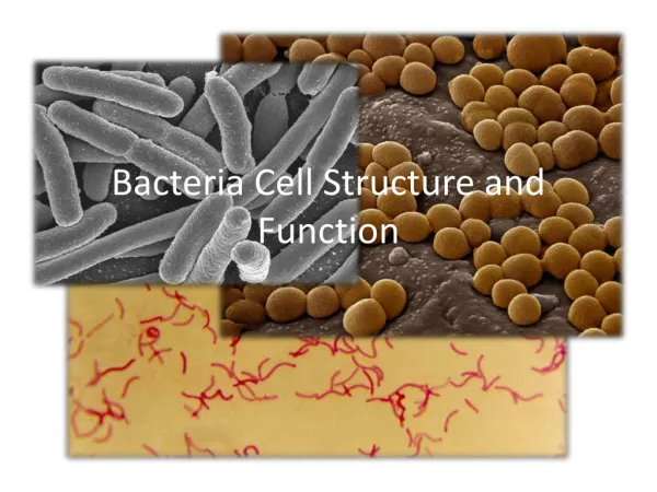
Bacteria constitute a large domain or kingdom of prokaryotic microorganisms • They were among the first life forms to appear on Earth, and are present in most habitats on the planet According to one of the researchers, "You can find microbes everywhere — they're extremely adaptable to conditions, and survive wherever they are”
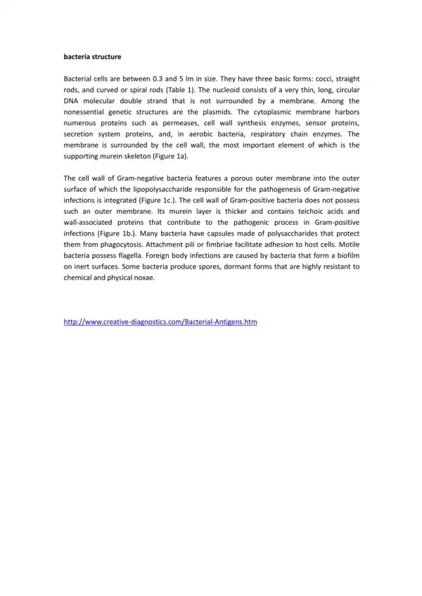
Size of Bacteria SIZE OF BACTERIA • Bacteria range in size from about 0.2 to 5 μ m • The smallest bacteria (Mycoplasma) are about the same size as the largest viruses (poxviruses), and are the smallest organisms capable of existing outside a host • The longest bacteria rods are the size of some yeasts and human red blood cells (7 μ m)
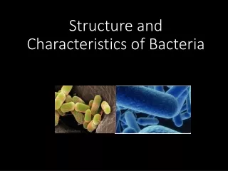
Shapes of Bacteria • Bacteria are classified by shape into three basic groups: • Cocciround • Bacilli rods • Spirochetes spiral shaped • Some bacteria are variable in shape and are said to be pleomorphic (many-shaped)
Bacterial Structures • Flagella • Pili • Capsule • Plasma Membrane • Cytoplasm • Cell Wall • Lipopolysaccharides • Teichoic Acids • Inclusions • Spores
Cell Wall • Outermost component common to all bacteria • Multilayered structure located external to the cytoplasmic membrane • Composed of an inner layer of peptidoglycan and an outer membrane that varies in thickness and chemical composition depending upon the bacterial type
The cell wall has several other important properties: • In gram-negative bacteria, it contains endotoxin, a lipopolysaccharide • Polysaccharides and proteins are antigens useful in laboratory identification • Porin proteins play a role in facilitating the passage of small, hydrophilic molecules into the cell
Peptidoglycan • The term "peptidoglycan" - peptides and the sugars (glycan) that make up the molecule • A complex, interwoven network that surrounds the entire cell • Composed of a single covalently linked macromolecule • Found only in bacterial cell walls • Provides rigid support for the cell, maintaining the characteristic shape of the cell, and allows the cell to withstand media of low osmotic pressure, such as water
Peptidoglycan structure: Escherichia coli (A) has a different cross-link from that of Staphylococcus aureus (B). In E. coli, c is cross-linked directly to d, whereas in S. aureus, c and d are cross-linked by five glycines. However, in both organisms the terminal D-alanine is part of the linkage. M, muramic acid; G, glucosamine; a, L-alanine; b, D-glutamic acid; c, diaminopimelic acid (A) or L-lysine (B); d, D-alanine; x, pentaglycine bridge.
Lipopolysaccharide • Lipopolysaccharide (LPS) of the outer membrane of the cell wall of gram-negative bacteria is endotoxin • Responsible for fever and shock (especially hypotension) • Called endotoxin because it is an integral part of the cell wall, in contrast to exotoxins, which are actively secreted from the bacteria • The LPS is composed of three distinct units : • A phospholipid called lipid A, which is responsible for the toxic effects • A core polysaccharide of five sugars linked through ketodeoxyoctulonate (KDO) to lipid A • An outer polysaccharide consisting of up to 25 repeating units of three to five sugars This outer polymer is the important somatic, or O, antigen of several gram-negative bacteria that is used to identify certain organisms in the clinical laboratory
Teichoic Acid • Fibers of glycerol phosphate or ribitol phosphate are located in the outer layer of the gram-positive cell wall • Some polymers of glycerol teichoic acid penetrate the peptidoglycan layer and are covalently linked to the lipid in the cytoplasmic membrane, called lipoteichoic acid • Ability to induce septic shock when caused by certain gram-positive bacteria • Mediate the attachment of staphylococci to mucosal cells. Gram-negative bacteria do not have teichoic acids
CYTOPLASMIC MEMBRANE • Just inside the peptidoglycan layer of the cell wall lies the cytoplasmic membrane • Composed of a phospholipid bilayer • The membrane has four important functions: • active transport of molecules into the cell • energy generation by oxidative phosphorylation • synthesis of precursors of the cell wall • secretion of enzymes and toxins
CYTOPLASM • The cytoplasm has two distinct areas • An amorphous matrix • ribosomes, nutrient granules, metabolites, and plasmids • A nucleoid region • composed of DNA
Ribosomes • Site of protein synthesis as in eukaryotic cells • 70S in size, with 50S and 30S subunits • Whereas eukaryotic ribosomes are 80S in size, with 60S and 40S subunits • Granules • Serve as storage areas for nutrients and stain characteristically with certain dyes • For example, volutin is a reserve of high energy stored in the form of polymerized metaphosphate, appears as a "metachromatic" granule since it stains red with methylene blue dye instead of blue • Metachromatic granules - Corynebacteriumdiphtheriae (diphtheria)
Nucleoid • The area of the cytoplasm in which DNA is located • DNA of prokaryotes is a single, circular molecule • Contains no nuclear membrane, no nucleolus, no mitotic spindle, and no histones • Bacterial DNA has no introns, whereas eukaryotic DNAdoes
Plasmids • Double-stranded • Circular DNA molecules • Extrachromosomal • Plasmids occur in both gram-positive and gram-negative bacteria, and several different types of plasmids can exist in one cell: • Transmissible plasmids • transferred from cell to cell by conjugation • large (MW 40–100 million) • they contain about a dozen genes responsible for synthesis of the sex pilus and for the enzymes required for transfer • Nontransmissible plasmids • are small (MW 3–20 million) • they do not contain the transfer genes
Plasmids carry the genes for the following functions and structures of medical importance: Antibiotic resistance Resistance to heavy metals such as mercury and silver, which is mediated by a reductase enzyme Resistance to ultraviolet light, which is mediated by DNA repair enzymes Pili (fimbriae), which mediate the adherence of bacteria to epithelial cells Exotoxins, including several enterotoxins
Transposons • Pieces of DNA that move readily from one site to another - jumping genes • Code for drug-resistant enzymes, toxins, or a variety of metabolic enzymes • Transposons typically have four identifiable domains
Specialized Structures Outside the Cell Wall • Capsule • Gelatinous layer covering the entire bacterium • Composed of polysaccharide • Sugar components of the polysaccharide vary from one species of bacteria to another • The capsule is important for four reasons: • Determinant of virulence of many bacteria since it limits the ability of phagocytes to engulf the bacteria • Specific identification of an organism can be made by using antiserum against the capsular polysaccharide called the quellung reaction • Capsular polysaccharides are used as the antigens in certain vaccines • Play a role in the adherence of bacteria to human tissues, which is an important initial step in causing infection
Flagella • Long, whiplike appendages that move the bacteria toward nutrients and other attractants, a process called chemotaxis • Flagellated bacteria have a characteristic number and location of flagella: • some bacteria have one, and others have many • in some, the flagella are located at one end, in others, they are all over the outer surface • Only certain bacteria have flagella
Pili (Fimbriae) • Hairlike filaments that extend from the cell surface • Shorter and straighter than flagella • Composed of subunits of pilin, a protein arranged in helical strands • Found mainly on gram-negative organisms • Pili have two important roles: • Mediate the attachment of bacteria to specific receptors • Role of s- pilus, during conjugation
Glycocalyx (Slime Layer) • A polysaccharide coating secreted by many bacteria • Covers surfaces like a film and allows the bacteria to adhere firmly to various structures, e.g., Skin, heart valves, and catheters • Important component of biofilms • Glycocalyx-producing strains of Pseudomonas aeruginosa, which cause respiratory tract infections in cystic fibrosis patients • Staphylococcus epidermidis and viridans streptococci – endocarditis • Streptococcus mutans, adherence to the surface of teeth-formation of plaque, the precursor of dental caries
SPORES • Highly resistant structures formed in response to adverse conditions • Bacillus-anthrax, • Clostridium-tetanus and botulism • Spore formation (sporulation) occurs when nutrients, such as sources of carbon and nitrogen, are depleted • Importance of spores lies in their extraordinary resistance to heat and chemicals. As a result of their resistance to heat, sterilization cannot be achieved by boiling Steam heating under pressure (autoclaving) at 121°C, usually for 15-20 minutes, is required to ensure the sterility of products for medical use
CELL WALLS OF GRAM-POSITIVE AND GRAM-NEGATIVE BACTERIA The structure, chemical composition, and thickness of the cell wall differ in gram-positive and gram-negative bacteria • The peptidoglycan layer is much thicker in gram-positive than in gram-negative bacteria • Some gram-positive bacteria also have fibers of teichoic acid, which protrude outside the peptidoglycan, whereas gram-negative bacteria do not • Gram-negative bacteria have a complex outer layer consisting of lipopolysaccharide, lipoprotein, and phospholipid • Between the outer-membrane layer and the cytoplasmic membrane in gram-negative bacteria is the periplasmic space, which is the site, in some species, of enzymes called -lactamases that degrade penicillins and other -lactam drugs
Comparison of Cell Walls of Gram-Positive and Gram-Negative Bacteria