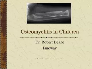
Chronic osteomyelitis
520 likes | 956 Views
Chronic osteomyelitis. Done by:Mohammad Al- M omani. Chronic osteomyelitis.

Chronic osteomyelitis
E N D
Presentation Transcript
Chronic osteomyelitis Done by:Mohammad Al-Momani
Chronic osteomyelitis • chronic osteomyelitis is a severe, persistent, and sometimes incapacitating infection of bone and bone marrow. It is often a recurring condition because it is difficult to treat definitively. A common sequel to acute haematogenous osteomyelitis. • An area of bone has been • destroyed by the acute • infection leaving sequestra • surrounded by dense sclerosed • bone called involucrum • sequestraprovoke a chronic seropurulent • Discharge which escapes through a sinus • (or several sinuses) at the skin surface.
causes • Bacteria can remain dormant for years,giving rise to recurrent acute flares and purulent discharges. • The usual suspects are • S. aureus, E. coli, S. pyogenes, Proteus and Pseudomonas
Sign and symptoms • Following acute bone infection, the patient returns with
Diagnosis • Sinogram(Culture of the discharge): • can help to localize the focus of active infection, • however, that a superficial swab sample may not reflect the really persistent infection; samples should be taken from deeper tissues. • Bonescans: • are useful in revealing hidden foci of inflammatory activity. • x-ray: • features of bone rarefaction • surrounded by dense sclerosis and • cortical thickening; within that area • there may be an obvious sequestrum.
CT and MRI: • are invaluable in planning operative • treatment: • together they will show the extent • of bone destruction and reactive • oedema, hidden abscesses • and sequestra • Technetium-99m diphosphonate bone scanning • (MDP) bone scans are usually positive 24 hours after an acute infection, and the scans demonstrate a well-defined focus of tracer activity 1-2 hours after the injection.
Treatment • Treatment depends on the frequency of relapsing flare-ups. • if seldom, it can be conservative. • if an abscess presents it should be incised. • Sequestrectomy should be performed only if a sequestrum is radiologically visible and surgically accessible.
conservativly • Antibiotics • Chronic infection is seldom eradicated by antibiotics alone. But the drug must be capable of penetrating sclerotic bone and should be non-toxic with long-term use. • (a) to suppress the infection and prevent its spread to healthy bone • (b) to control acute flares • Antibiotics are administered for 4–6 weeks (starting from the beginning of treatment or the last debridement) before considering operative treatment. • Local treatment • A sinus may be painless and need dressing simply to protect the clothing. An acute abscess may need urgent incision and drainage.
operation • External fixation: • may need to be applied so that internal fixation devices can be removed. • All infected and dead tissue must then be excised. After 3 or 4 days the wound is inspected and if there are renewed signs of tissue death the debridement is repeated – several times if necessary. Antibiotic cover is continued for at least 4 weeks after the last debridement.
complications • pathologic fracture • secondary amyloidosis • endocarditis • sepsis • development of squamous cell carcinoma in the draining sinus tracts • sarcoma in the infected bone.
Septic arthritis is inflammation of a synovial membrane with purulent effusion into the joint cavity, due toinfection. • The joint is invaded through a penetrating wound, by eruption of an adjacent bone abscess or by blood spread from a distant site. As infection spreads through the joint, articular cartilage is eroded; • Synovialmembrane • Membrane surroundingjoint cavity • Produce synovial fluid • Contain rich capillary network for phagocytic and hyaluronate- producingfunction
Bacterial, but sometimes viral,mycobacterial, andfungal. • Usually caused byStaphylococcus aureus . Other organisms are : E.coli , Proteus , Streptococcus • Predisposing Factor: • Rheumatoidarthritis • Immunosuppressive drugtherapy • Age >80 or very young children • AIDS • Chronicdisorder • Intravenous drugabuse
Pathogenesis • Bacteria can gain entrance to a joint via manyroutes:
Most common form ofspread Usually affect people with underlying medicalproblem May result from penetratingtrauma Introduction of organisms during diagnostic and surgical procedures. For egarthroscopy and intra-articularinjection More common inchildren. Osteomyelitisusually begin in the metaphyseal region, from whichit breaks through the periosteuminto the joint.
Synovial membrane is highlyvascularised. ↓ Bacteria can easily enter synovial joint via bloodstream. ↓ There will be inflammatory reaction with seropurulent exudate and increase in synovialfluid. ↓ As pus appear in the joint, the articular cartilage is eroded and destroyed. Partly by the bacterial enzyme, and partly by the enzyme released from synovium, inflammatory cell and pus Adult Infant Destroy theepiphysis, which is still largely cartilaginous. Children Vascular occlusion lead to necrosis of epiphysealbone Effect confined on articular cartilage Extensive erosioncan occur due to synovial proliferation and ingrowth
a) In the early stage, there is an acute synovitis with a purulent joint effusion • b) Soon the articular cartilage is attacked by bacterial and cellular enzyme. • c) If infection is not arrested , the cartilage may be completely destroyed • d) Healing then leads to ankylosis
If left untreated, it will spread to the underlying bone and out of joint to form abscess andsinus. \ • Healingwith: • Completeresolution • Partial loss of articular cartilage and fibrosis of joint • 3.Loss of articular cartilage and bony ankylosis • 4.Bony destruction and permanentdeformity
Inchildren • acute pain in single large joint • (esphip) Inadults • Often in the superficial joint(knee, wrist or ankle) • Joints painful, swollen • &inflamed. • Pseudoparesis • Child is ill,rapid pulse andswingingfever • Warmth andmarked local tenderness &movementrestricted. • Overlying skin looks red & superficial joint swelling may be obvious • look for gonococcal infection or drugabuse. • Local warmth and markedtenderness • Patient with rheumatoid arthritisand especially those on corticosteroidmay • develop “silent” joint infection. • All movements are restricted by pain orspasm. • Look for source of infection from septic toe or dischargeear
Synovial fluidanalysis • Aseptic technique is used during • aspiration of synovialfluid. • Avoid taken from infected site of • skin. • The fluid is then analyzed by • gross and microscopic • examination andculture. • Gross examinations include appearance, volume, viscosity, mucin clotting (amount ofproteoglycans). • Microscopic examinations include leucocytecount, stainingof smears, serum glucose ratio,protein. • Finally, culture and sensitivitytestfor definitivediagnosisandtreatment.
Imaging • Xray • Early Stage – Normal except widening of joint space, ultrasoundhelpful • Look for soft tissue swelling, loss of tissue planes, widening of joint space and slight subluxation due to fluid in joint. Gas may be seen with E. coli infection • Late stage – Narrowing and irregularity of jointspace • Plain film findings of superimposed osteomyelitis may develop • (periosteal reaction, bone destruction, sequestrumformation). • MRI and radionuclide imaging • are helpful in diagnosing arthritis in obscuresites such as the sacroiliac and sterno-clavicularjoint.
subchondral erosionsand Joint spaceloss sclerosis of the femoral head
Treatment General supportivecare -Analgesics -IVfluids Splintage - The joint must be rested either on a splint or in a widely split plaster -In neonates and infants, with hip infection the joint is held abducted and 30 degree flexed, on traction to prevent dislocation. Antibiotics Treatment is started once the blood and samples are obtained without waiting for the detailresults. Choice of antibiotic depends on the most likelypathogen
SurgicalManagement • Arthrocentesis-a needle is placed into the joint to extract ad remove thejoint fluid. • Arthroscopicdebridement and copious irrigation with normal saline – more frequently in knee joint septicarthritis • Amputation esp. in ischemia &risk of sepsis
DIFFERENTIALDIAGNOSIS • Acuteosteomyelitis • Trauma • Hemophilicbleed • Rheumaticfever • Juvenile rheumatoidarthritis • Sickle-celldisease • Gaucher’sdisease • Gout andpseudo-gout
Complications • Bonedestructionanddislocation of the joint (esp Hip) • Cartilage destruction • -may lead to either fibrosis or bonyankylosis • - in adult partial destruction of the joint will result in secondaryosteoarthritis • Growth disturbance • - presenting as either localised deformity or shortening of thebone