Download
1 / 108
1.08k likes | 1.31k Views
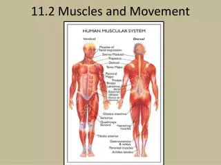
Cellular movement and Muscles. Muscles – general information. Vertebrates and many invertebrates have three main classes of muscle Skeletal muscle connect bones are are used for complex coordianted activities.

E N D
Muscles – general information Vertebrates and many invertebrates have three main classes of muscle • Skeletal muscle connect bones are are used for complex coordianted activities. • Smooth musclessurround internal organs such as the large and small intestines, the uterus, and large blood vessels • The contraction and relaxation of smooth muscles controls the diameter of blood vessels and also propels food along the gastrointestinal tract. • Compared with skeletal muscles, smooth muscle cells contract and relax slowly, and they can create and maintain tension for long periods of time. • Cardiac muscle: Striated muscle of the heart.
Muscles - introduction • B. Skeletal muscle from the neck of a hamster • C. Heart muscle from a rat • D. Smooth muscle from the urinary bladder of a guinea pig • E. Myoepithelial cells in a secretory alveolus from a lactating rat mammary gland
Microtubule Function • Move subcellular components • Use motor proteins kinesin and dynein • e.g., Rapid change in skin color
Microtubules Show Dynamic Instability • Balance between growth and shrinkage • Factors • Local concentrations of tubulin • Dynamic instability • Microtubule-associated proteins (MAPs) • Temperature • Chemicals can disrupt the dynamics (e.g., plant poisons)
Movement Along Microtubules • Direction is determined by polarity and the type of motor protein • Kinesin move in + direction • Dynein moves in – direction • Fueled by ATP • Rate of movement is determined by the ATPase domain of the protein and regulatory proteins • Dynein is larger than kinesin and moves 5-times faster
Cilia and Flagella • Cilia – numerous, wavelike motion • Flagella – single or in pairs, whiplike movement • Composed of microtubules • Arranged into axoneme • Movement results from asymmetric activation of dynein
Microfilaments • Other type of cytoskeletal fiber • Polymers composed of the protein actin • Often use the motor protein myosin • Found in all eukaryotic cells • Movement arises from • Actin polymerization • Sliding filament model using myosin (more common)
Microfilament Structure and Growth • Polymers of G-actin called F-actin • Spontaneous growth (6-10X faster at + end) • Treadmilling when length is constant • Capping proteins increase length by stabilizing minus end
Actin Polymerization • Amoeboid movement • Two types • Filapodia are rodlike extensions • Neural connections • Microvilli of digestive epithelia • Lamellapodia resemble pseudopodia • Leukocytes • Macrophages
Skeletal muscle (striated muscle) • Skeletal muscle cells are one of the largest cells in the body • Are multinucleate formed by the fusion of myoblasts • Diameters range from 50 to 150 microns with lengths ranging from mm to cm • Muscle fibers contract in response to an electrical signal ie depolarization • The signal is generated at the synapse (the neuromuscular junction) and propagated through an action potential via the muscle fiber membrane • The membrane of the cell has specialized invaginations called Transverse-tubules (T-tubules) that enter into the cell (at every 1-2 microns) • The action potential can be rapidly transmitted deep into the interior of the cell resulting a delay of only 3-5 msec between the depolarization at the synapse and the first muscle fiber tension. • The T-tubule network is so extensive that 50-80% of the plasma membrane is in the T-tubules.
Neuromuscular junction – things to remember • Each muscle fiber is innervated by a motorneuron • One motorneuron can innervate one or multiple fibers • Each motorneuron plus its complement of muscle fibers is called a motor unit as well all contract in concert. • The synpase between the motorneuron and the muscle fiber is called a neuromuscular junction(NMJ) • Nerve terminal contains many mitochondria and vesicles which can be seen lined up in double rows along side the voltage-gated Ca2+ channels attached to presynaptic membrane => active zone. • The vesicles of the NMJ have very high concentrations of neurotransmitter (2,000 to 10,000 molecules of ACH per vesicle) • Excess of neurotransmitter is released to ensure that the resulting post-synaptic depolarization is strong enough to generate an action potential - "safety margin"
The nACHR - again • The receptor is made up of 4 different transmembrane proteins one of which (the alpha subunit) is repeated to give 5 subunits to create the ion channel • ACH binds to the alpha subunit and thus it takes two molecules of acetylcholine to open the channel • One nACHR opens and allow 1.5 x 10⁴ Na+ ions/msec of open time • The channel opens on average 1 msec. • 1 vesicle contains enough neurotransmitter to open ~3000 receptors (wow!) and because two molecules of Ach is needed to open one receptor there must be a minimum of ~6,000 molecules Ach per vesicle. • Studies have shown that the amount of neurotransmitter contained in one vesicle causes an post-synaptic potential of ~ 1 mV. • If the average depolarization generated at a NMJ • of a muscle fiber is 40 mV then there must be at • least 40 vesicles released and in the order of • 120,000 receptors activated at the NMJ. WOW!
Striated muscle channels and action potentials • Pumps and transporters • 1) Na+/K+ ATPase pump - to establish the electrochemical gradients of Na+ and K+ • 2) Ca2+ ATPase pump - uses energy from ATP to remove 2 Ca2+ from the inside to the outside of the cell to ensure that internal Ca2+ concentrations remain low (10-7 mM internal) • 3) Na+/Ca2+ cotransporter - to also remove Ca2+ from the inside of the cell and uses the energy from the cotransport of 3 Na+ molecules to export 1 Ca2+ • 4) Muscle Ca2+ ATPase pump - a different pump from number 2 above. Found highly concentrated on the sarcoplasmic reticulum (SR) (constitutes 80% of the protein found in the SR membranes). • The muscle Ca2+ ATPase pumps 2 Ca2+ into the SR to lower cytosolic Ca 2+ and to concentrate Ca 2+ into internal stores.
Striated muscle channels and action potentials Channels - muscle cells share many of the same ion channels as neurons • Leak channels - besides the leak K+ channel, skeletal muscle cells have a high concentration of Cl- leak channels. The high permeability to Cl- helps repolarize the membrane after an action potential 2) Voltage-gated Na+ channels . 3) Voltage-gated K+ channel - the delayed rectifier K+ 4) Voltage gated Ca2+channels - high threshold Ca2+ channels
Skeletal muscle (striated muscle) • Terminology • Muscle cell: Muscle fiber • Myofibrils: Main intracellular structures in striated muscles. Are bundles of contractile and elastic proteins • Sarcolemma: Cell membrane of a muscle cell • Cytoplasm: Sarcoplasm • Sarcoplasmic reticulum: wraps around each myofibril like a piece of lace. Release and sequester Ca2+ ions
Skeletal muscle (striated muscle) • The sarcoplasmic reticulum (SR) regulates the cytosolic Ca2+ levels in skeletal muscle • Myofibril: A long bundle of actin, myosin and associated proteins in muscle cell. • Transverse (T) tubules: invaginations of the plasma membrane, enter • myofibers at the Z disks, where they come in close contact with the • terminal cisternae of the SR • Terminal cisternae: store Ca2+ ions and connect with the lacelike • network of SR tubules that overlie the A band.
T-Tubules and SRs • Transverse tubules • Sarcolemmal invaginations • Enhance action potential penetration • More developed in larger, faster twitching muscles • Sarcoplasmic reticulum (SR) • Stores Ca2+ • Terminal cisternae - storage
Triads and Sarcoplasmic reticulum • The link between depolarization and Ca2+ release or excitation-contraction coupling occurs at the junctions between the T-tubule and the sarcoplasmic reticulum • 80% of the T-tubules membrane is associated with the sarcoplasmic reticulum at triads • The voltage-gated Ca2+ channels are concentrated in the T-tubules in the triads • The Ca2+ release channel found in the sarcoplasmic reticulum membrane is associated with the voltage-gated Ca2+ channel at this point • This close association allows for the rapid signaling from action potential to Ca2+ release.
General sequence of events • Resting [Ca2+]i = 0.1 μM • AP propagation along Sarcolemma and into T- tubules • Depolarization opens the voltage-gated Ca2+ channels at triad junctions • This results in a release of Ca2+ through the Ca2+ release channels from the SR • Cytosolic [Ca2+]i reaches 1-10 μM • Diffusion and binding of Ca2+ to TnC • Contraction events • [Ca2+] to resting levels: • 1. After the action potential is passed and the voltage-gated Ca2+ channels close, the Ca2+ release channels close • 2. Ca2+ is recycled back into the SR through the Ca2+ ATPases • 3. Ca2+ binds to calsequesterin
Ca2+ channels and its release • Release of Ca2+ stores mediated by ryanodine receptors (RYRs) in skeletal muscle • Voltage sensing dihydropyridine (DHP) receptors in the plasma membrane contact ryanodine receptors located in the membrane of the SR • In response to a change in voltage, the dihydropyridine receptors undergo a conformational change • This produces a conformational change in the associated RYRs, opening them so that Ca2+ ions can exit into the cytosol. • The voltage-gated Ca2+ channel is either closely localized to or makes a physical connection to the Ca2+ release channels in the SR • Cont……..
Ca2+ channels and its release • Not all Ca2+ release channels are associated with voltage-gated Ca2+ channels • These non-associated channels are thought to be opened solely by Ca2 influx into the cytosol from the voltage-gated Ca2+ channels. • The Ca2+ release channel in the SR of most muscle cells (smooth, cardiac, skeletal) is a Ca2+ activated Ca2+ channel • The Ca2+ release channel is stimulated to open at low concentrations of Ca2+ ( up to 0.1 mM) in the cytosol but inhibited by high concentrations of Ca2+ in the cytosol (0.5 mM and higher for cardiac cells) • So as Ca2+ is released from the SR it starts to inhibit the Ca2+ release channel.
Sarcomeres • Skeletal muscle is made up of bundles of multinucleate muscle cells (myofibers) • Each cell contains myofibrils that are composed of repeated units of actin and myosin called sacromeres • Thick and thin filaments arranged into sarcomeres • Repeated in parallel and • in series • Features • Z-disk • A-band • I-band • M-lines
Sarcomeres • Electron micrograph of a longitudinal section through a skeletal muscle cell of a rabbit • Schematic diagram of a single sarcomere • Z discs: At each end of the sarcomere • Attachment sites for the plus ends of actin filaments (thin filaments) • M line: Midline. • Location of proteins that link adjacent myosin II filaments (thick filaments) to one another • Dark bands: mark the location of the thick filaments = A bands • Light bands: which contain only thin filaments and therefore have a lower density of protein = I bands.
Myofibril • A single continuous stretch of interconnected sarcomeres • Runs the length of the muscle cell • More myofibrils in parallel more force
Striated Muscle Cell Structure • Composed of thick and thin filaments • Thick: myosin • 300 myosin II hexamers • Thin: actin • Capped by tropomodulin (-) and CapZ (+) to stabilize • Decorated by troponin and tropomyosin • Globular protein (G-actin) • Form long chains called F-actin • In skeletal muscle 2 F-actin polymers twist together
Myosin • Motor protein used by actin • Sliding filament model • Most common type of movement • Myosin is an ATPase • Converts E released • from ATP to mechanical E • 17 classes of myosin with • multiple isoforms • Similar structure • Head, tail, and neck
Sliding Filament Model • Analogous to pulling yourself along a rope • Actin: the rope • Myosin: your arm
Sliding Filament Model • Two processes • Chemical • Myosin binds to actin (Cross-bridge) • Structural • Myosin bends (Power stroke) • Cross-bridge cycle • Formation of cross-bridge, power stroke, and release • Need ATP to attach and release • No ATP rigor mortis
Sliding Filament Model, Cont. • Myosin is bound to actin in the absence of ATP and this is the "rigor" state i.e. gives rigidity to the muscle • ATP binds to the myosin causing the head domain to dissociate from actin • ATP is then hydrolyzed causing a conformational change in the mysoin head to move it to a new position and bind to actin • Pi is released causing the myosin head to change conformation again and it is this movement that moves the actin • ADP is released
Ca2+ Allows Myosin to Bind to Actin • Ca2+ binds to TnC • Reorganization of troponin-tropomyosin • Expose myosin-binding site on actin
Ca2+ Allows Myosin to Bind to Actin * * * * * • Ca2+ levels increase in cytosol • Ca2+ binds to troponin C • Troponin-Ca2+ complex pulls tropomyosin away form G-actin binging site • Myosin binds to actin and completes power stroke • Actin filament moves
Contractile Force • Depends on sarcomere length: distance between the Z-disks • Number of myofibrils • Number of cells (recruitment)
Regulation of Contraction • Excitation-contraction coupling • Depolarization of the muscle plasma membrane (sarcolemma) • Elevation of intracellular Ca2+ • Contraction • Relaxation when the sarcolemma repolarizes and Ca2+ returns to resting levels
Excitation – contraction coupling • ACH released at the NMJ • Net entry of Na+ initiates a muscle action potential • AP in T- tubule alters conformation of DHP receptor • DHP receptor opens Ca2+ release channels in SR and Ca2+ enters the cytoplasm • Ca2+ binds to troponin C • Troponin-Ca2+ complex pulls tropomyosin away form G-actin binging site • Myosin binds to actin and completes power stroke • Actin filament moves
Cause of Depolarization • Myogenic • Spontaneous • e.g., Vertebrate heart • Pacemaker • Cells that depolarize fastest • Unstable resting membrane potential • Neurogenic • Excited by neurotransmitters • e.g., Vertebrate skeletal muscle • Can have multiple (tonic) or single (twitch) innervation sites