Download
1 / 1
10 likes | 104 Views
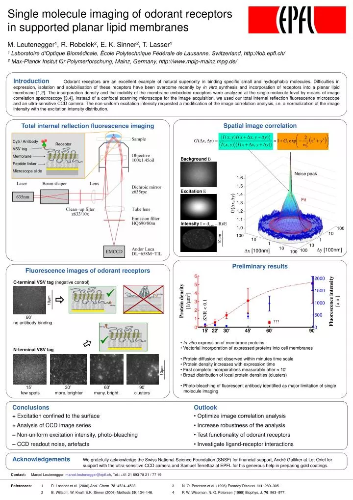
Cy5 / Antibody VSV tag Membrane Peptide linker Microscope slide. Receptor. Noise peak. 1.6 1.5 1.4 1.3 1.2 1.1 1.0. Fit. G ( D x , D y ). 100. 10. 100. 10. 10 m m. 1. 1. 10. 10. D y [100nm]. D x [100nm]. 100. 100. 10 m m. 10 m m.

E N D
Cy5 / Antibody VSV tag Membrane Peptide linker Microscope slide Receptor Noise peak 1.6 1.5 1.4 1.3 1.2 1.1 1.0 Fit G(Dx,Dy) 100 10 100 10 10mm 1 1 10 10 Dy [100nm] Dx [100nm] 100 100 10mm 10mm Introduction Odorant receptors are an excellent example of natural superiority in binding specific small and hydrophobic molecules. Difficulties in expression, isolation and solubilisation of these receptors have been overcome recently by in vitro synthesis and incorporation of receptors into a planar lipid membrane [1,2]. The incorporation density and the mobility of the membrane embedded receptors were analyzed at the single-molecule level by means of image correlation spectroscopy [3,4]. Instead of a confocal scanning microscope for the image acquisition, we used our total internal reflection fluorescence microscope and an ultra-sensitive CCD camera. The non-uniform excitation intensity requested a modification of the image correlation analysis, i.e. a normalization of the image intensity with the excitation intensity distribution. Single molecule imaging of odorant receptors in supported planar lipid membranes Total internal reflection fluorescence imaging Spatial image correlation BackgroundB ExcitationE IntensityI = (Iraw – B)/E • Preliminary results • In vitro expression of membrane proteins • Vectorial incorporation of expressed proteins into cell membranes • Protein diffusion not observed within minutes time scale • Protein density increases with expression time • First complete incorporations measurable after 10’ • Broad distribution of local protein densities (clusters) • Photo-bleaching of fluorescent antibody identified as major limitation of singlemolecule imaging M. Leutenegger1, R. Robelek2, E. K. Sinner2, T. Lasser1 1 Laboratoire d‘Optique Biomédicale, École Polytechnique Fédérale de Lausanne, Switzerland, http://lob.epfl.ch/ 2Max-Planck Insitut für Polymerforschung, Mainz, Germany, http://www.mpip-mainz.mpg.de/ Fluorescence images of odorant receptors C-terminal VSV tag (negative control) 60’ no antibody binding N-terminal VSV tag 15’ 30’ 60’ 90’ few spots more, brighter many, bright clusters Protein density[1/mm2] Fluorescence intensity[a.u.] SNR < 0.1 ??? Conclusions Outlook + Excitation confined to the surface • Optimize image correlation analysis + Analysis of CCD image series • Increase robustness of the analysis – Non-uniform excitation intensity, photo-bleaching• Test functionality of odorant receptors – CCD readout noise, artefacts • Investigate ligand-receptor interactions AcknowledgementsWe gratefully acknowledge the Swiss National Science Foundation (SNSF) for financial support, André Galliker at Lot-Oriel for support with the ultra-sensitive CCD camera and Samuel Terrettaz at EPFL for his generous help in preparing gold coatings. Contact: Marcel Leutenegger, marcel.leutenegger@epfl.ch, Tel.: +41 21 693 78 21 / 77 19 References: 1 D. Lossner et al. (2006) Anal. Chem. 78: 4524–4533. 3 N. O. Petersen et al. (1998) Faraday Discuss. 111: 289–305. 2 B. Wiltschi, W. Knoll, E.K. Sinner (2006) Methods 39: 134–146. 4 P. W. Wiseman, N. O. Petersen (1999) Biophys. J. 76: 963–977.