Download

1 / 14
140 likes | 558 Views
Understand the exfoliation patterns and different cell types in urine cytology, including urothelial, squamous, prostatic, and renal cells. Learn to identify normal and reactive cells, dysplasia, metaplastic cells, and their clinical significance.

E N D
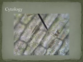
1 Urine Cytology Soheir Mahfouz 2017
NORMAL CELLS Soheir Mahfouz -Self Study Series
LOW Cellularity Single cells Clear background NORMAL SMEAR 1- PATTERN OF EXFOLIATION (LOW CELLULARITY clean) Single cell exfoliation: Mainly urothelial cells in small numbers. The number depends on the method. More cells in cystoscopic samples, bladder washing or after exercise In groups: Usually seen after catheterization or after cystoscopy 2-CELL TYPES • Urothelial cells • Superficial /umbrella cells • Intermediate • Deep cells • Squamous cells: exfoliate from distal urethra in male or are vulvo-vaginal in females. They may also exfoliate in the presence of stones-parasites-trichomonas • Prostatic cells & Seminal vesicle cells : rare except after prostatic massage • Renal cells: rare • WBC should be less than 5/HPF if more consider urinary tract inflammation & RBCs should be less than 1/several HPF Soheir Mahfouz -Self Study Series
NB : A-crystals: normally should not be present B-casts: important in the follow up of renal transplants for rejection/ indicate renal disease • Hyaline: acceptable 1 or 2 in a smear*. They are pale blue homogenous, well defined & cylindrical transparent, may have orange colour if increased it means excess protein filtration in nephrotic / nephritic syndromes • Cellular/epithelial: normal none or 1/10 HPF, they are desquamated cell + protein. If they contain fat patient is diabetic • Granular casts may be found after strenuous exercise otherwise pathologic NB: if the casts are wide in caliber (tubular dilatation ) bad sign. • WBC: with sharp contours, indicates: pyelonephritis • RBC casts: normally not present They indicate hemorrhagiccondition in kidneys: AGN- polyarteritisnodosa-crush syndrome, hepatorenal A.Graft rejection NB: Pseudocasts are trapped WBC with irregular contour and originate from lower urinary tract infection C-Fungus(alternaria)-vorticells (protozoa of fresh water) – parasites- organisms in water or plant material pollen Soheir Mahfouz -Self Study Series
Candida Ova in a pseudocast Renal cast Corpora amylacea HPV Alternaria Polyoma virus Soheir Mahfouz -Self Study Series
Normal urothelial cells 1) Umbrella cells (largest) 2) Intermediate cells, 3) Basal cells (smallest). Conspicuous nucleoli are only seen in intermediate and basal urothelial cells, in bladder washings. The nuclear membranes are smooth and the cells have abundant vacuolated(bubbly) cytoplasm. 4)+/- a benign columnar cell, from the bladder dome or trigone. Soheir Mahfouz -Self Study Series
Umbrella cells Normal umbrella cells. Surface urothelial cells covering the underlying cells • Abundantpear shaped cytoplasm and low N/C ratio. • Thickened outer cytoplasmic margins ("saucer cup edge") and vacuolatedperinuclear areas. • nuclei are uniform in size and central- bi & multinucleation common Soheir Mahfouz -Self Study Series
Umbrella cells Soheir Mahfouz -Self Study Series
Reactive cells Soheir Mahfouz -Self Study Series
Reactive cells • Reactive urothelial cells with distinctive nucleoli and multiple cytoplasmic vacuoles N.B • Use the lymphocyte and squamous cell for size comparisons. • Sheets are monolayer DD TCC umbrella squamous Soheir Mahfouz -Self Study Series
Dysplasia (UIN) • NB: ALL KERATINIZING dysplasia are considered high grade / CIS Sheets monolayer - no nucleolus- No mass Dysplasia may be seen in both transitional & squamous epithelial cells: The cells show irregularity in shape & size of nuclei. There is noticeable irregularity of chromatin density & pattern - Mild dysplasia / low grade UIN is insignificant clinically - Severe dysplasia requires cystoscopic biopsy and follow up DD TCC - CIS : Single cell exfoliation- NO MASS – No tumour diathesis-NO nucleolus ( DD HG TCC) Soheir Mahfouz -Self Study Series
Metaplastic cells Metaplastic smears ARE GENERALLY CELLULAR ONES displaying inflammatory changes A) Squamous cells: 1-cytoplasm pink –orange with double contour 2-Nucleus may be vesicular ( mature metaplasia) B) Columnar cells: uniform cells with basal nuclei Soheir Mahfouz -Self Study Series
Columnar metaplasia & UIN Soheir Mahfouz -Self Study Series