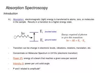
LIGHT ABSORPTION SPECTROSCOPY
LIGHT ABSORPTION SPECTROSCOPY. colorimetric analysis of nmol samples of macromolecules Prof. Eric Wickstrom. E. B. orthogonal electronic (E) and magnetic (B) components of linearly polarized light. Prism Slit.

LIGHT ABSORPTION SPECTROSCOPY
E N D
Presentation Transcript
LIGHT ABSORPTION SPECTROSCOPY colorimetric analysis of nmol samples of macromolecules Prof. Eric Wickstrom
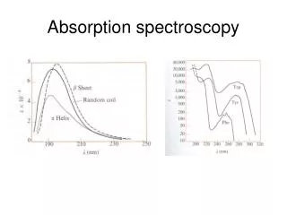
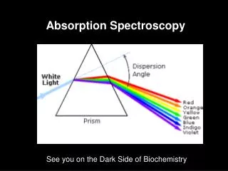
E B orthogonal electronic (E) and magnetic (B) components of linearly polarized light
Prism Slit
I0 is the current measured by the photomultiplier tube when the light goes through the reference cuvet with buffer but not sample. • I is the current measured by the photomultiplier tube when the light goes through the complete sample. • The ratio of I to I0 is called the transmittance, T, so T = I/I0 . Therefore T ranges from 0 (total sample absorbance) to 1 (no absorbance at all).
The absorbance, A, is defined as A= -log T. • Because it is a logarithm, absorbance has no units. • When the sample absorbs 90% of the light beam, then T = 0.1 = 1/10, and A = -log(1/10) = +log(10) = 1 • When the sample absorbs 99% of the light beam, then T = 0.01 = 1/100, and A = -log(1/100) = +log(100) = 2
The most commonly used unit is the absorbance unit, AU, or OD in biochemical slang, an amount of a substance that will give an absorbance of 1.0 at the wavelength of interest in a cuvet with a pathlength of 1.0 cm, when dissolved in 1.0 mL of buffer. 1 mg DNA = 20 A260U 1 mg protein = 1 A280U
We write the relationship as A= εlc, where l is the pathlength in cm, and c is the concentration in mole/liter. • The extinction coefficient, or molar absorptivity, ε, is the theoretical absorbance of a 1.0 M (mole/liter) solution in 1.0 cm cuvet. • Because absorbance is unitless, ε has units of liter/mole·cm.
Typical Initial Concentrations • Protein Concentration: 0.5 mg/mL • Cell Path Length: 10 mm • Stabilizers (Metal ions, etc.): minimum • Buffer Concentration : 5 mM or as low as possible while maintaining protein stability • Contaminants: Unfolded protein, peptides, particulate matter (scattering particles)