Download
1 / 8
160 likes | 779 Views
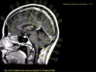
4. 5. 6. 7. 8. 9. 1. 2. 3. 11. 12. 10. 13. 24. 14. Identify anatomical structures 1 - 24. 18. 19. 17. 20. 21. 16. 22. 15. 23. Fig. 1.10 A midline Post-contrast Sagittal T 1 Weighted MRI. 4. 5. 6. 7. 8. 9. 1. 2. 3. 11. 12. 10. 13. 24. 14. 1. Scalp fat

E N D
4 5 6 7 8 9 1 2 3 11 12 10 13 24 14 Identify anatomical structures 1 - 24 18 19 17 20 21 16 22 15 23 Fig. 1.10 A midline Post-contrast Sagittal T1 Weighted MRI
4 5 6 7 8 9 1 2 3 11 12 10 13 24 14 1. Scalp fat 2. Bone 3. Inferior sagittal sinus 4. Corpus callosum 5. Internal cerebral vein 6. Vein of Galen 7. Superior sagittal sinus 8. Parietal lobe 9. Occipital lobe 10. Straight sinus 11. Vermis 12. IV ventricle 13. Cerebellar tonsil 14. Cervical cord 15. Medulla 16. Pons 17. Midbrain 18. Mass intermedia of thalamus 19. Anterior III ventricle 20. Optic chiasm 21. Pituitary gland 22. Sphenoid sinus 23. Nasopharynx 24. Frontal lobe 18 19 17 20 21 16 22 15 23 Fig. 1.10 A midline Post-contrast Sagittal T1 Weighted MRI
8 7 6 5 4 2 3 1 Identify anatomical structures 1 - 8 Fig. 1.11. Coronal Section of the Brain at the level of IV Ventricle Post Contrast Coronal T1 Weighted MRI
8 7 6 5 4 2 3 1 1. Cerebellar tonsil 2. Cerebellar hemisphere 3. IV ventricle 4. Superior vermis 5. Tentorium 6. Posterior temporal lobe 7. Choroid plexus within lateral ventricle 8. Posterior frontal lobe Fig. 1.11. Coronal Section of the Brain at the level of IV Ventricle Post Contrast Coronal T1 Weighted MRI
1 5 6 7 9 2 3 4 8 11 10 Identify anatomical structures 1 - 12 12 Fig. 1.12. Coronal Section of the Brain at the level of Pituitary gland Post Contrast Coronal T1 Weighted MRI
1 5 6 7 9 2 3 4 1. Frontal lobe 2. Corpus callosum 3. Frontal horn 4. Caudate nucleus 5. III ventricle 6. Optic nerve 7. Pituitary stalk 8. Pituitary gland 9. Internal carotid artery 10. Cavernous sinus 11. Sphenoid sinus 12. Nasopharynx 8 11 10 12 Fig. 1.12. Coronal Section of the Brain at the level of Pituitary gland Post Contrast Coronal T1 Weighted MRI
1 2 3 4 5 Identify anatomical structures 1 - 5 Fig. 1.13. Coronal Section of the Brain at the level of the orbits. Post Contrast Coronal T1 Weighted MRI.
1 2 3 4 5 1. Frontal lobe 2. Orbital Fat 3. Globe 4. Nasal Cavity 5. Maxillary Sinus Fig. 1.13. Coronal Section of the Brain at the level of the orbits. Post Contrast Coronal T1 Weighted MRI.