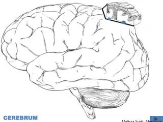
CEREBRUM
CEREBRUM. Dr. Zeenat Zaidi Dr. Essam Eldin Salama. Objectives. At the end of the lecture, the student should be able to: List the parts of the cerebral hemisphere (cortex, medulla, basal nuclei, lateral ventricle). Describe the subdivision of a cerebral hemisphere into lobes.

CEREBRUM
E N D
Presentation Transcript
CEREBRUM Dr. Zeenat Zaidi Dr. Essam Eldin Salama
Objectives At the end of the lecture, the student should be able to: • List the parts of the cerebral hemisphere (cortex, medulla, basal nuclei, lateral ventricle). • Describe the subdivision of a cerebral hemisphere into lobes. • List the important sulci and gyri of each lobe. • Describe different types of fibers in cerebral medulla (association, projection and commissural) and give example of each type.
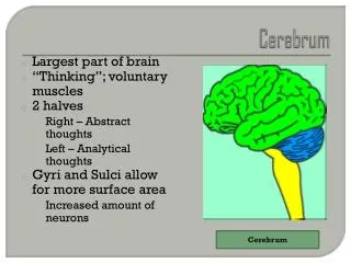
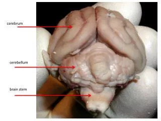
Cerebrum Corpus callosum • Largest part of the forebrain. • Divided into two halves, the cerebral hemipheres, whichare separated by a deep median longitudinal fissure which lodges the falx cerebri. • In the depth of the fissure, the hemispheres are connected by a bundle of fibers called the corpus callosum. Left hemisphere Right hemisphere Median longitudinal fissure
Surfaces Superolateral Medial Inferior (tentorial)
Includes: Cerebral cortex: Superficial layer of grey matter White matter (WM): Deeper to the cortex, containing axons to and from the cells of the cortex Basal ganglia:Number of nuclear masses buried within the white matter Lateral ventricle: The cavity of hemisphere Structure of Cerebrum Cortex Basal ganglia WM WM Lateral ventricle
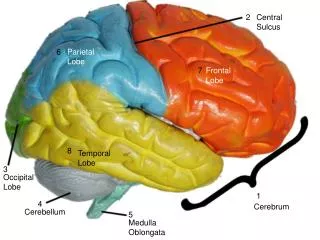
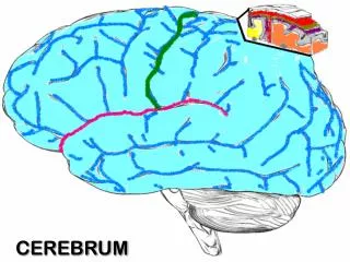
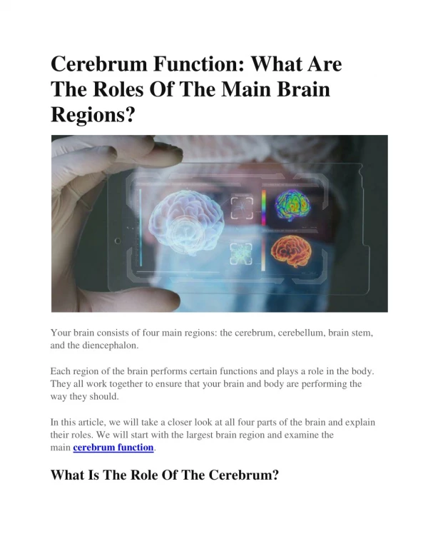
Lobes of Cerebrum The superficial layer of grey matter is highly convoluted to form a complex pattern of ridges (gyri) and grooves (sulci). This arrangement maximizes the surface area of the cerebral cortex (about 70% is hidden within the depths of sulci). • Three sulci, consistent in position, named central, lateral (sylvian) & parieto-occipital, divide each hemisphere into FOUR lobes: Frontal, Parietal, Temporal & Occipital (named after overlying bones) by • Functionally each hemisphere contains a ‘limbic lobe’ on the medial surface. S g
Function of Each Lobe reception and evaluation of sensory information motor function, motivation, aggression, smell and mood visual processing emotions, memory storage & Linking conscious intellectual functions with the unconscious autonomic functions, smell, hearing, memory and abstract thought
Frontal lobe: Precentralgyrus. Superior & inferior frontal sulci divide the lobe into superior, middle & inferior frontal gyri. • Parietal lobe: • Postcentralgyrus. • Intraparietalsulcusdividing the lobe into superior & inferior parietal lobules. Precentralgyrus Postcentralgyrus sfs Superior parietal lobule ifs Inferior parietal lobule Intraparietal sulcus Superior , middle & inferior frontal gyri
Temporal lobe: Superior & inferior temporal sulci giving rise to superior, middle & inferior temporal gyri. Insula: the gyri in the depth of lateral fissure, covered by parts of frontal, parietal & temporal lobes called the opercula (removed in lower picture.). Superior, middle & inferior temporal gyri sts its insula
Medial Surface • Sulci: Parietooccipital, Calcarine, Cingulate • Gyri: Cingulate, Parahippocampal
Brodmann’s Map • Brodmann produced a numbered, cytological map of cerebral cortex based upon its regional histological characteristics • Subdivisions with similar cellular and laminar structure are called 'areas' • Brodmann's numbering of these cortical locations has become one of the standard ways to identify brain areas.
Frontal Lobe Premotor cortex: Located in the region immediately anterior to the precentral gyrus (Brodmann’s area 6). Primary motor cortex:Located in precentral gyrus (Brodmann area 4). Prefrontal cortex: Extensive region of the frontal lobe anterior to premotor area. Broca’s (motor speech) area: Located in the inferior frontal gyrus of the dominant hemisphere, usually left (Brodmann’s area 44 & 45). Frontal eye field: Located in the middle frontal gyrus immediately in front of motor cortex (Brodmann’s area 8).
Parietal lobe Primary somatosensory cortex: located in postcentral gyrus (Brodmann’s area 1, 2, 3). Parietal association cortex: located posterior to primary somatosensory cortex. Occipital lobe • Primary visual cortex: located on the medial surface of the hemisphere, in the gyri surrounding the calcarinesulcus(Brodmann’s area 17). • Visual association cortex: located around the primary visual cortex.
Primary auditory cortex: located in the superior surface of the superior temporal gyrus (Brodmann’s area 41, 42) Temporal Lobe Auditory association cortex: located immediately around the primary auditory cortex (also includes Wernick’s area) Parahippocampalgyrus:located in the inferomedial part of temporal lobe. Deep to this gyrus lies the hippocampus and the amygdala, which are parts of limbic system
Language Area • Organized around the lateral fissure. • Broca’s area: concerned with expressive aspects of language. • Wernick’s area: responsible for comprehension of the spoken words. • Nearby regions of temporal lobe and parietal lobe (angular gyrus & supramarginal gyrus of the inferior parietal lobule) are important in naming, reading, writing, and calculation.
Hemispheric Dominance • The localization of speech centers & mathematical ability is the criterion for defining the dominant cerebral hemisphere. • In 96% of normal right-handed individuals and 70% of normal left-handed individuals, the left hemisphere contains the language centers. These are left hemisphere dominant. • Cerebral dominance becomes established during the first few years after birth. Verbal Memory Shape Memory Hemispheres communicate via the corpus callosum
White Matter • Underlies the cortex, contains nerve fibers, neuroglia cells and blood vessels. • The nerve fibers originate, terminate or sometimes both, within the cortex. • Depending on their origin & termination, these nerve fibers are classified into three types: Association, Projection & Commissural Association fibers: Unite different parts of the same hemisphere, are of two types: long & short Commissural fibers:Connect the corresponding regions of the two hemispheres Projection fibers: Consist of afferent and efferent fibers of the cerebral cortex
Short association fibersconnect adjacent gyri, Long association fibers connect more distant parts and include: Uncinate fasciculus: connects frontal to temporal lobe Superior longitudinal fasciculus: connects the frontal, occipital, parietal, and temporal lobes Arcuate fasciculus: connect gyri in frontal to temporal lobes Inferior longitudinal fasciculus: connects occipital to temporal pole Cingulum: connects frontal & parietal lobes to the para-hippocampalgyrus and adjacent temporal gyri Association Fibers Short association fibers 2 5 3 1 4
Commissural Fibers 1 3 • Connect the corresponding regions of the two hemispheres. • Include: • Corpus callosum. • Anterior commissure. • Posterior commissure. • Hippocampal commissure (commissure of fornix). 2
Corpus Callosum Anterior forceps • Connects the corresponding regions of the two hemispheres except the temporal lobes, that are connected by anterior commissure • It is shorter craniocaudally than is the hemisphere • The callosal fibers linking the frontal poles curve forward forming anterior forceps (forceps minor) • The callosal fibers linking the occipital poles curve backward forming posterior forceps (forceps major) F C C P O Posterior forceps
Parts of Corpus Callosum Genu Splenium Body Rostrum
Posterior Commissure: connects the left and right midbrain. Important in the bilateral pupillary reflex Corpus callosum • Anterior commissure: connects the inferior and middle temporal gyri & the olfactory regions of the two hemispheres Thalamus Midbrain • Hippocampal Commissure: connects the two hippocampi with each other
Projection Fibers corona radiata • Consist of Afferent & Efferent of the cerebral cortex. • Deeper to the cortex, these fibers are arranged radially as the corona radiata. • Then the fibers converge downward, form internal capsule, between thalamus and basal ganglia. • Continue in the crus cerebri of the midbrain, basilar part of pons, & pyramid of medulla oblongata. Internal capsule crus cerebri pyramid pyramidal decussation
Internal Capsule • Bundle of projection fibers, passes through the interval between the thalamus and the basal ganglia • Has 5 parts: • Anterior limb: Thalamocortical & Frontopontine fibers • Genu:corticobulbar fibers • Posterior limb: Corticospinal, Corticobulbar & Thalamocortical fibers • Retrolenticular part: Geniculocalcarine fibers • Sublenticular part (not shown):geniculo-temporal fibers C 1 2 L 3 T 4