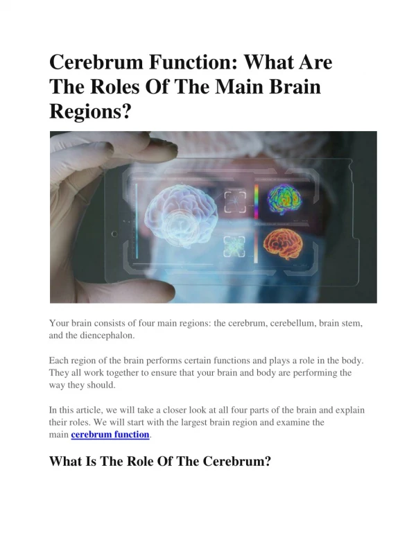
The Cerebrum
The Cerebrum . Lobes, the Cerebral Cortex, and Cortical Regions of the Brain. Cerebrum. Cerebrum. Cerebellum. Cerebrum - The largest division of the brain. It is divided into two hemispheres, each of which is divided into four lobes.

The Cerebrum
E N D
Presentation Transcript
The Cerebrum Lobes, the Cerebral Cortex, and Cortical Regions of the Brain
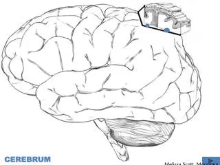
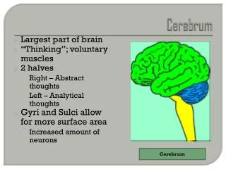
Cerebrum Cerebrum Cerebellum Cerebrum -The largest division of the brain. It is divided into two hemispheres, each of which is divided into four lobes. http://williamcalvin.com/BrainForAllSeasons/img/bonoboLH-humanLH-viaTWD.gif
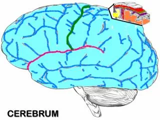
Cerebral Cortex CerebralCortex Cerebral Cortex - The outermost layer of gray matter making up the superficial aspect of the cerebrum. http://www.bioon.com/book/biology/whole/image/1/1-6.tif.jpg
Gyri – Elevated ridges “winding” around the brain. • Sulci – Small grooves dividing the gyri Cerebral Features: • Central Sulcus – Divides the Frontal Lobe from the Parietal Lobe • Fissures – Deep grooves, generally dividing large regions/lobes of the brain • Longitudinal Fissure – Divides the two Cerebral Hemispheres • Transverse Fissure – Separates the Cerebrum from the Cerebellum • Sylvian/Lateral Fissure – Divides the Temporal Lobe from the Frontal and Parietal Lobes
Gyri – Elevated ridges “winding” around the brain. • Sulci – Small grooves dividing the gyri Cerebral Features: • Central Sulcus – Divides the Frontal Lobe from the Parietal Lobe • Fissures – Deep grooves, generally dividing large regions/lobes of the brain • Longitudinal Fissure – Divides the two Cerebral Hemispheres • Transverse Fissure – Separates the Cerebrum from the Cerebellum • Sylvian/Lateral Fissure – Divides the Temporal Lobe from the Frontal and Parietal Lobes
Specific Sulci/Fissures: Central Sulcus Longitudinal Fissure Sylvian/Lateral Fissure Transverse Fissure http://www.bioon.com/book/biology/whole/image/1/1-8.tif.jpg http://www.dalbsoutss.eq.edu.au/Sheepbrains_Me/human_brain.gif
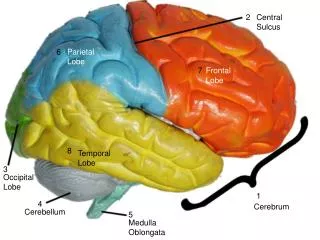
Lobes of the Brain (4) • Frontal • Parietal • Occipital • Temporal http://www.bioon.com/book/biology/whole/image/1/1-8.tif.jpg * Note: Occasionally, the Insula is considered the fifth lobe. It is located deep to the Temporal Lobe.
Lobes of the Brain - Frontal • The Frontal Lobe of the brain is located deep to the Frontal Bone of the skull. • It plays an integral role in the following functions/actions: • - Memory Formation • - Emotions • - Decision Making/Reasoning • - Personality (Investigation: Phineas Gage) Investigation (Phineas Gage) Modified from: http://www.bioon.com/book/biology/whole/image/1/1-8.tif.jpg
Frontal Lobe - Cortical Regions • Primary Motor Cortex (Precentral Gyrus) – Cortical site involved with controlling movements of the body. • Orbitofrontal Cortex – Site of Frontal Lobotomies • Broca’s Area – Controls facial neurons, speech, and language comprehension. Located on Left Frontal Lobe. • Broca’s Aphasia – Results in the ability to comprehend speech, but the decreased motor ability (or inability) to speak and form words. * Possible Side Effects: - Epilepsy - Poor Emotional Responses - Perseveration (Uncontrolled, repetitive actions, gestures, or words) • * Desired Effects: • - Diminished Rage • - Decreased Aggression • - Poor Emotional Responses • Olfactory Bulb - Cranial Nerve I, Responsible for sensation of Smell
Lobes of the Brain - Parietal Lobe • The Parietal Lobe of the brain is located deep to the Parietal Bone of the skull. • It plays a major role in the following functions/actions: - Senses and integrates sensation(s) • Spatial awareness and perception • (Proprioception - Awareness of body/ body parts in space and in relation to each other) Modified from: http://www.bioon.com/book/biology/whole/image/1/1-8.tif.jpg
Parietal Lobe - Cortical Regions • Primary Somatosensory Cortex (Postcentral Gyrus) – Site involved with processing of tactile and proprioceptive information. • Somatosensory Association Cortex - Assists with the integration and interpretation of sensations relative to body position and orientation in space. May assist with visuo-motor coordination. • Primary Gustatory Cortex – Primary site involved with the interpretation of the sensation of Taste.
Lobes of the Brain – Occipital Lobe • The Occipital Lobe of the Brain is located deep to the Occipital Bone of the Skull. • Its primary function is the processing, integration, interpretation, etc. of VISION and visual stimuli. Modified from: http://www.bioon.com/book/biology/whole/image/1/1-8.tif.jpg
Occipital Lobe – Cortical Regions • Primary Visual Cortex – This is the primary area of the brain responsible for sight -recognition of size, color, light, motion, dimensions, etc. • Visual Association Area – Interprets information acquired through the primary visual cortex.
Primary Visual Cortex Visual Association Area Regons Modified from: http://www.bioon.com/book/biology/whole/image/1/1-8.tif.jpg
Lobes of the Brain – Temporal Lobe • The Temporal Lobes are located on the sides of the brain, deep to the Temporal Bones of the skull. • They play an integral role in the following functions: • Hearing • Organization/Comprehensionof language • Information Retrieval (Memory and Memory Formation) Modified from: http://www.bioon.com/book/biology/whole/image/1/1-8.tif.jpg
Temporal Lobe – Cortical Regions • Primary Auditory Cortex – Responsible for hearing • Primary Olfactory Cortex – Interprets the sense of smell once it reaches the cortex via the olfactory bulbs. (Not visible on the superficial cortex) • Wernicke’s Area – Language comprehension. Located on the Left Temporal Lobe. - Wernicke’s Aphasia – Language comprehension is inhibited. Words and sentences are not clearly understood, and sentence formation may be inhibited or non-sensical.
Lobes and Structures of the Brain A. G. B. F. C. E. D. http://williamcalvin.com/BrainForAllSeasons/img/bonoboLH-humanLH-viaTWD.gif
Lobes and Structures of the Brain A. Central Sulcus B. Frontal Lobe C. Sylvian/Lateral Fissure A. (groove) G. D. Temporal Lobe B. F. E. Transverse Fissure F. Occipital Lobe C. (groove) G. Parietal Lobe E. D. (groove) http://williamcalvin.com/BrainForAllSeasons/img/bonoboLH-humanLH-viaTWD.gif
Further Investigation Phineas Gage: Phineas Gage was a railroad worker in the 19th century living in Cavendish, Vermont. One of his jobs was to set off explosive charges in large rock in order to break them into smaller pieces. On one of these instances, the detonation occurred prior to his expectations, resulting in a 42 inch long, 1.2 inch wide, metal rod to be blown right up through his skull and out the top. The rod entered his skull below his left cheek bone and exited after passing through the anterior frontal lobe of his brain. Frontal
Remarkably, Gage never lost consciousness, or quickly regained it (there is still some debate), suffered little to no pain, and was awake and alert when he reached a doctor approximately 45 minutes later. He had a normal pulse and normal vision, and following a short period of rest, returned to work several days later. However, he was not unaffected by this accident. http://www.sruweb.com/~walsh/gage5.jpg Learn more about Phineas Gage: http://en.wikipedia.org/wiki/Phineas_Gage Frontal