Download
1 / 65
670 likes | 1.06k Views
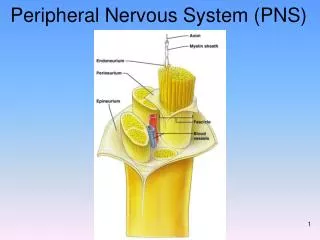
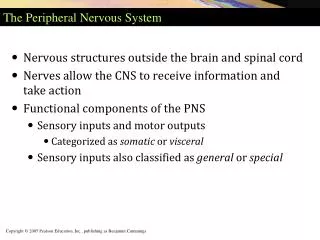
The Peripheral Nervous System (PNS). P A R T 2. Spinal Nerves. Thirty-one pairs of mixed nerves arise from the spinal cord and supply all parts of the body except the head They are named according to their point of issue 8 cervical (C 1 -C 8 ) 12 thoracic (T 1 -T 12 )

E N D
The Peripheral Nervous System (PNS) P A R T 2
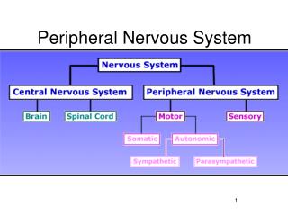
Spinal Nerves • Thirty-one pairs of mixed nerves arise from the spinal cord and supply all parts of the body except the head • They are named according to their point of issue • 8 cervical (C1-C8) • 12 thoracic (T1-T12) • 5 Lumbar (L1-L5) • 5 Sacral (S1-S5) • 1 Coccygeal (C0)
Spinal Nerves Figure 13.6
Spinal Nerves: Roots • Each spinal nerve connects to the spinal cord via two medial roots • Each root forms a series of rootlets that attach to the spinal cord • Ventral roots arise from the anterior horn and contain motor (efferent) fibers • Dorsal roots arise from sensory neurons in the dorsal root ganglion and contain sensory (afferent) fibers
Spinal Nerves: Roots Figure 13.7a
Spinal Nerves: Rami • The short spinal nerves branch into three or four mixed, distal rami • Small dorsal ramus • Larger ventral ramus • Tiny meningeal branch • Rami communicantes at the base of the ventral rami in the thoracic region • visceral nerve fibers
Nerve Plexuses • All ventral rami except T2-T12 form interlacing nerve networks called plexuses • Plexuses are found in the cervical, brachial, lumbar, and sacral regions • Each resulting branch of a plexus contains fibers from several spinal nerves
Nerve Plexuses • Each muscle receives a nerve supply from more than one spinal nerve • Damage to one spinal segment cannot completely paralyze a muscle
Spinal Nerve Innervation: Back, Anterolateral Thorax, and Abdominal Wall • The back is innervated by dorsal rami via several branches • The thorax is innervated by ventral rami T1-T12 as intercostal nerves • Intercostal nerves supply muscles of the ribs, anterolateral thorax, and abdominal wall
Spinal Nerve Innervation: Back, Anterolateral Thorax, and Abdominal Wall Figure 13.7b
Cervical Plexus • The cervical plexus is formed by ventral rami of C1-C4 • Most branches are cutaneous nerves of the neck, ear, back of head, and shoulders • The most important nerve of this plexus is the phrenic nerve • Motor and sensory nerve of the diaphragm
Cervical Plexus Figure 13.8
Brachial Plexus • Formed by C5-C8 and T1 (C4 and T2 may also contribute to this plexus) • It gives rise to the nerves that innervate the upper limb
Brachial Plexus • There are four major branches of this plexus (C5-T1) • Roots • Trunks • Divisions • Cords
Brachial Plexus Figure 13.9a
Brachial Plexus: Nerves • Axillary • Musculocutaneous • Median • Ulnar • Radial
Brachial Plexus: Distribution of Nerves Figure 13.9c
Brachial Plexus: Nerves Figure 13.9b
Lumbar Plexus • Arises from L1-L4 and innervates the thigh, abdominal wall, and psoas muscle • The major nerves are the • Femoral • For anterior thigh muscles • Obturator • Adductors muscles
Lumbar Plexus Figure 13.10
Sacral Plexus • Arises from L4-S4 and serves the buttock, lower limb, pelvic structures, and the perineum (pudendal nerve) • The major nerve is the sciatic, the longest and thickest nerve of the body • Lower limb (except anteromedial thigh muscles) • Branches into two nerves: the tibial and the common fibular (peroneal)
Sacral Plexus Figure 13.11
Dermatomes • A dermatome is the area of skin innervated by the cutaneous branches of a single spinal nerve • All spinal nerves except C1 participate in dermatomes
Dermatomes Figure 13.12
Innervation of Joints • Hilton’s law: any nerve serving a muscle that produces movement at a joint also innervates the joint itself and the skin over the joint
Motor Endings • PNS elements that activate effectors by releasing neurotransmitters at: • Neuromuscular junctions • Varicosities at smooth muscle and glands
Levels of Motor Control • The three levels of motor control are • Segmental level • Spinal cord circuit • Projection level • Pyramidal and extrapyramidal systems • Precommand level • Cerebellum and basal nuclei
Hierarchy of Motor Control Figure 13.13
Segmental Level • The segmental level is the lowest level of motor hierarchy • It consists of segmental circuits of the spinal cord • Its circuits control locomotion and specific, oft-repeated motor activity
Projection Level • Controls the spinal cord • Consists of: • Cortical motor areas that produce the direct (pyramidal) system • Brain stem motor areas that oversee the indirect (multineuronal) system • Send information to lower motor neurons and also to higher center
Precommand Level • Cerebellar and basal nuclei systems that: • Regulate motor activity • Precisely start or stop movements • Coordinate movements with posture • Block unwanted movements • Monitor muscle tone • Control the output of the cortex and brain stem motor centers
Reflexes • A reflex is a rapid, predictable motor response to a stimulus • Reflexes may: • Be inborn (intrinsic) or learned (acquired) • Involve, peripheral nerves, brain stem and spinal cord • Somatic and visceral reflexes
Reflex Arc • There are five components of a reflex arc • Receptor • Sensory neuron • Integration center • Motor neuron • Effector
Reflex Arc Figure 13.14
Somatic Reflexes • Spinal: • Stretch reflex • Golgi tendon reflex • Withdrawal reflex • Crossed-extensor reflex • Superficial: • Plantar • Babinski’s • Abdominal
Stretch and Deep Tendon Reflexes • For skeletal muscles to perform normally: • The Golgi tendon organs (proprioceptors) must constantly inform the brain as to the state of the muscle • Stretch reflexes initiated by muscle spindles must maintain healthy muscle tone
Stretch reflex - monosynaptic • Muscle Spindle • Are composed of intrafusal muscle fibers that lack myofilaments in their central regions, are noncontractile, and serve as receptive surfaces • Two types of afferent endings: • Primary sensory endings of type Ia fibers (faster conduction) • Secondary sensory endings of type II fibers (slower conduction)
Stretch reflex • Intrafusal fivers are innervated by gamma () efferent fibers • Extrafusal fibers • Contractile muscle fibers • Innervated by alpha () efferent fibers
Muscle Spindles Figure 13.15
Operation of the Muscle Spindles • Stretching the muscles activates the muscle spindle • There is an increased rate of action potential in Ia fibers • Contracting the muscle reduces tension on the muscle spindle • There is a decreased rate of action potential on Ia fibers
Figure 13-4b Muscle spindles monitor muscle length and prevent over-stretching Musclelength Action potentials in spindlesensory neuron Muscle returnsto initial length. Muscleis stretched. Time (1) (2) Increasedefferent outputthroughalpha motorneurons Increasedafferent signalsto spinalcord Firing rateof afferentsensoryneurondecreases Musclecontracts Spinalcord Musclestretch Negativefeedback (b) Muscle stretch can trigger a stretch reflex, which contracts themuscle to avoid over-stretching.
Operation of the Muscle Spindle Figure 13.17
Stretch Reflex - monosynaptic • Stretching the muscle activates the muscle spindle • Excited α motor neurons causes the muscle to contract • Afferent impulses from the spindle result in inhibition of the antagonist • Example: patellar reflex • Tapping the patellar tendon stretches the quadriceps and starts the reflex action • The quadriceps contract and the antagonistic hamstrings relax
Stretch Reflex Figure 13.16
Golgi Tendon Reflex - polysynaptic • The opposite of the stretch reflex • Contracting the muscle activates the Golgi tendon organs • Afferent Golgi tendon neurons are stimulated, neurons inhibit the contracting muscle, and the antagonistic muscle is activated • As a result, the contracting muscle relaxes and the antagonist contracts • It moderates the muscle contraction
Golgi Tendon Reflex The Golgi Tendon reflex: Reflex that causes muscle relaxation and lengthening in response to muscle contraction. Figure 13.18
Figure 13-6a–c Muscle reflexes help prevent damage to the muscle Muscle spindle reflex: the addition of a load stretches the muscle and the spindles, creating a reflex contraction. Sensory neuron Spindle Spinalcord Add load Motor neuron Muscle (a) Load added to muscle. (b) Muscle and muscle spindlestretch as arm extends. (c) Reflex contraction initiated by musclespindle restores arm position.
Figure 13-6d–e Muscle reflexes help prevent damage to the muscle Golgi tendon reflex protects the muscle from excessively heavy loads by causing the muscle to relax and drop the load. Inhibitinginterneuron 1 1 Musclecontracts Neuron from Golgi tendonorgan fires. 3 2 Motor neuron is inhibited. 2 Motor neuron Golgi tendonorgan 3 Muscle relaxes. 4 4 Load is dropped. (d) Muscle contraction stretchesGolgi tendon organ. (e) If excessiveload is placed onmuscle, Golgi tendonreflex causes relaxation,thus protecting muscle.
Flexor ( Withdrawal) Reflexes • The flexor reflex happens on the limb receiving the painful stimulus • Withdrawal reflex by contraction of the flexor muscles • Reciprocal inhibition of the extensors • Polysynaptic reflex