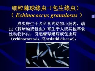
echinococcus
echinococcus u044du0445u0438u043du043eu043au043eu043au043a

echinococcus
E N D
Presentation Transcript
CLASSIFICATION Kingdom : Animalia Phylum : Platyhelminthes Class : Cestoda Order : Cyclophyllidea Family : Taenidae Genus : Echinococcus Species : granulosus
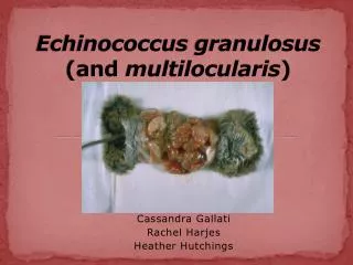
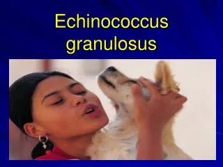
INTRODUCTION • Echinococcus granulosus, also called hydatid worm belongs to class Cestoda • It causes cystic echinococcosis in livestock and humans being intermediate hosts and parasitize the small intestines of adult canids • It is a zoonotic disease • Definitive hosts are carnivorous predators like dogs, wolves, foxes and lions. While sheep, goat, cattle, pigs and rodents are intermediate hosts. Birds and arthropods act as mechanical vectors
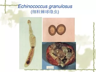
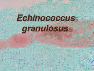
MORPHOLOGY • The adult tapeworm ranges in length from 2 mm to 7mm and has three proglottids when intact — a immature proglottid, mature proglottid and a gravid proglottid. • It has scolex with four suckers andalso has a rostellum withhooks. • Echinococcus is triploblastic, anus is absent and it has no digestivesystem. • Its body is covered by tegument andthe worm is divided into a scolex, a short neck, and three to six proglottids. Its body shape isribbon-like.
TRANSMISSION • Adult E. granulosus release eggs within the intestine which will be transported out of the body via feces • When contaminated waste is excreted into the environment, intermediate host has the potential to contract the parasite by grazing in contaminated pasture • It is transmitted from the intermediate host (sheep) to the definitive host (dogs) by frequent feeding of offal. • Consuming offal containing Echinococcus granulosus can lead to infection
LIFE CYCLE • The adult is in the small intestines of the definitive host (dogs) • Gravid proglottids release eggs that are passed in the feces • The intermediate hosts are infected by ingesting eggs, the egg hatches in the small bowel and releases an oncosphere • The oncosphere penetrates the intestinal wall and moves through the circulatory system to various organs • In the organs they develop into cysts and enlarge gradually
The cysts produce protoscolices and daughter cysts • Definitive host eats the infected organs and becomes infected • After ingestion, the protoscolices evaginate, attach to the intestinal mucosa and develop into adult stages • In 32-80 days, cycle starts over
The growth rate of cystsis highly variable and may depend on strain differences and cyst location. Estimates of the average increase of cyst diameter vary (approximately 1.5-2 cm/year).
Gross pathology of hydatid daughter cysts Pathologically hydatid liver cyst has three distinct layers: membrane and Ectocyst - fibrous adventitial layer due to host response Middle layer - laminated membrane of proteinaceous material Endocyst - inner germinal layer from which the scolices may be detached from human lung
A hydatid cyst in the cranium of a child (the ruler at the top measures 6 inches long, and the child's brain is below the hydatid cyst). This infection resulted in the child's death.
PATHOGENESIS • Ingested eggs from animal hatch in thegut and releaseoncospheres • Oncospheres penetrate the intestinal wall, migrate via the circulation, and lodge in the liver or lungs or, less frequently, in the brain, bone, or otherorgans. • In tissue , E. granulosus oncospheres develop into cysts, which grow slowly (usually over many years) into large fluid-filled lesions—hydatidcysts • Large cysts may contain >1 L of highly antigenic hydatid fluid as well as millions of protoscolices • If a cyst in the liver leaks or ruptures, infection can spread to theperitoneum.
SIGNS • Signs depend upon the site ofinfection • Liver cysts cause abdominal pain ofa palpable mass. Jaundice may occur if the bile duct isobstructed. • Rupture into the bile duct, peritoneal cavity, or lung may cause fever, urticaria, or a serious anaphylacticreaction. • Pulmonary cysts can causecough, chest pain, andhaemoptysis. • Brain and spinal cord; causeepilepsy and blindness
DIAGNOSIS • Diagnosis in the definitive host, the dog is difficult by ordinary microscopy as it cannot demarcate between Taenia and Echinococcus eggs • Detection of antigens in feces by ELISA is currently the best available technique Other techniques are; Imaging Serologic testing Examination of cyst fluid
TREATMENT • Surgical Removal of Hydatid Cysts 90% effective but can be risky depending on location, size, and advancement of cyst • It may need chemotherapy to prevent recurrance • Chemotherapy: Albendazole is preferred treatment because it penetrates into hydatid cysts. Dosage: 10mg/kg body weight or 400mg 2x daily for 4 weeks Mebendazole Dosage: 40mg/kg body weight 3x daily for 3- 6 months Dogs are effectively treated with Praziquental • PAIR Treatment Puncture, aspiration, injection, respiration • Inject protoscolicidal substances into the cyst
PREVENTION • In order to prevent transmission to dogs from intermediate hosts, dogs can be given anthelminthicvaccinations • Clean slaughter and high surveillance of potential intermediate host during slaughter is key in preventing the spread this cestode to its definitive host • Proper disposal of carcass and offal after slaughter to prevent dogs access to offal from livestock • Boiling livers and lungs which contain hydatid cysts for 30 minutes has been proposed as a simple, efficient and saving way to kill the infectiouslarvae