Download
1 / 25
340 likes | 859 Views
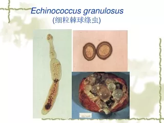
Echinococcus granulosus ( 细粒棘球绦虫 ). GENERAL INTRODUCTION Worldwide distribution Extra-intestinal tapeworm Small tapeworm Laval infection of E. granulosus may cause serious clinical disease ---hydatidosis/ hydatid disease. Morphology. Only 2-8 mm long Usually comprises of-

E N D
GENERAL INTRODUCTION • Worldwide distribution • Extra-intestinal tapeworm • Small tapeworm • Laval infection of E. granulosus maycause serious clinical disease ---hydatidosis/ hydatid disease
Morphology • Only 2-8 mm long • Usually comprises of- • Scolex: with four suckers and 2 circular rows of hooks • neck • immature proglottid • mature proglottid • gravid proglottid • The eggs of E.granulosus and Teania spp. are indistinguishable
Hydatid Cyst: • Round & cystic • Wall – cuticle layer, germinal layer • Contents • cystic fluid, brood capsules, protoscolex, daughter & grand daughter cysts (hydatid sands)
Hydatid cyst Cuticle layer Germinal layer Daughter cyst Granddaugher cyst Protoscolex Brood capsule Brood capsule
Definitive host: dog & other canine • Intermediate host: sheep, cattle, camel & human • Infective stage: egg (gravid proglottid) • Sites of hydatid:liver, lungs, abdominal cavity, spleen, kidneys, heart, bones, central nervous system etc • Man is a dead end host
Pathogenesis • Cause Hydatid Disease (Hydatidosis) • Sites of hydatid cyst: liver, lungs, abdominal cavity, spleen, kidney, heart, bones, brain etc Analysis of 15,289 cases in Xinjiang,China • Liver 69.97% • Lung 19.3% • Abdominal cavity 3%
Clinical menifestations • Depends on the size, the location and the number of cyst. • Pressure –by tremendous size of the cyst. results in disfunction of liver, lung or nervous system • Allergy -due to rupture of cyst, may cause severe allergic reaction • Regeneration – due to rupture of cyst, intracystic protoscolex or germinal layer may be transplanted and result in multiplesecondary infection Secondary regeneration 5.3% • Toxicosisby secretion of worm
Diagnosis • History of contacting with sheep & dogs • Clinical symptoms of a slow-growing tumor accompanied by eosinophilia are suggestive • Parasitological examination for finding scolexes, brood capsules & daughter cysts • Cysts in organs or calcified cysts can be visualized using x-rays, CT & B-ultrasound examination • Biopsy are forbidden unless during operation
Serological examination for specific Ab or Cag. • Intradermal (Casoni) test with hydatid fluid is useful. • Antibodies against hydatid fluid antigens have been detected in a sizable population of infected individuals by ELISA or indirect hemagglutination test.
CT, brain B-ultrasound, liver X-ray, lung CT, liver
Epidemiology World distribution. South America (Argentina, Brazil, Uruguay etc ),North America (American, Canada etc) Europe(Iceland, Russia, France, Spain etc), Africa (Kenya, Libya, Egypt, Tunisiaetc ).
Endemic Factors • Contamination of the feces by infected dogs • Intimate contact between dog, herbivorous animal and man in local district • Traveling to endemic areas & importing from endemic areas
Control and treatment • Regular treatment of infected dogs to reduce worm load. • Prevention of dogs from eating infected offals of domestic animals(sheep,etc) in the endemic areas. • Health education and strict personal hygiene. • Avoidance of unnecessary contact with infected dogs. • Surgery is still remains the mainstay of the treatment of hydatid disease. • Albendazole have proved to be effective against hydatid cyst(for median or small size cysts).
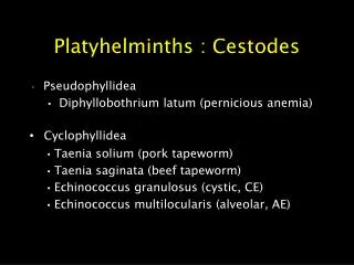
Summary T. solium T. saginata E. granulosus Proglottides700-1000 1000-2000 only 3 Rostellumpresent absent present Hooks 25-50 absent 24-48 Uterine branches 7-13 15-30 unregularly branched Definitive hosthuman human dog/wolf Intermediate hostpig/human cattle sheep/human Infective stageegg/cysticercus cysticercus egg