Pulmonary Echinococcus
Pulmonary Echinococcus. Megan Brundrett December 15, 2009. Pathology 1. Pathology 2. Echinococcus. Cestodes – divide their life cycle in 2 or more hosts Intermediate host – immature parasite as a tissue cyst Definitive host – mature parasite as a tapeworm Echinococcus granulosus

Pulmonary Echinococcus
E N D
Presentation Transcript
Pulmonary Echinococcus Megan Brundrett December 15, 2009
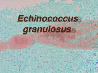
Echinococcus • Cestodes – divide their life cycle in 2 or more hosts • Intermediate host – immature parasite as a tissue cyst • Definitive host – mature parasite as a tapeworm • Echinococcus granulosus • Echinococcus multilocularis
E. granulosus • Hydatid disease • Definitive hosts are dogs or other canines • Intermediate hosts – sheep, goats, camels, horses, humans • Australia, New Zealand, Argentina, Chile, Ireland, Scotland, middle Europe, Kenya
E. multilocularis • Alveolar disease • Definitive host – foxes, and other canines • Intermediate host – rodents, humans • Arctic areas of United States, Canada, and Russia
Life Cycle • (1) Adult tapeworms in bowels of definitive host • (2) Eggs passed in feces, ingested by intermediate host • (3) Onchosphere penetrates intestinal wall, carried via blood vessels to lodge in organs • (4) Hydatid cysts develop • (5) Protoscolices (larvae) ingested by definitive host • (6) Attach to small intestine of definitive host and grow to adult worm.
Hydatid disease • E. granulosus • 75% hepatic involvement, 25% pulmonary • Other organs – brain, bone, heart, kidney • Cause symptoms with mass effect, rupture, secondary infection
Alveolar disease • E. multilocularis • < 5% of symptomatic Echinococcal infections • Lacks a limiting membrane like hydatid disease and causes more infiltration to surrounding tissues. • Clinical presentation – jaundice, RUQ pain, malaise • If untreated >90% die within 10 years • Now prognosis improved b/c of albendazole therapy. Very difficult to surgically resect.
Pulmonary Echinococcal Diease • 60% right lung • 50-60% in lower lobes • Lungs may be a more common site in children • 30% have multiple cysts in the lungs • 20% of pts with lung involvement have liver involvement
Clinical Manifestations • Unruptured cyst: Chest pain, hemoptysis, chronic cough • Ruptured cyst: Cough, fever, expectorating salty material (hydatid membrane and larvae). • Acute hypersensitivity with rupture: Fever, urticaria, sometimes anaphylaxis • Superinfection by bacteria, fungal presenting with fever, sepsis • Pulmonary HTN, Pulmonary embolism
Diagnosis • CXR: Round or oval mass with smooth borders. No calcifications. If pericyst (fibrous capsule that host makes) incorporates bronchioles, than air penetrates between the pericyst and exocyst creating a meniscus sign or crescent shape. • CT scan: Thin enhancing ring if cyst is intact. Fluid filled in center. Homogenous.
Differential Diagnosis • Primary lung cancer • Metastatic cancer • Tuberculosis • Bronchogenic Cyst • Round pneumonia • Aspergillosis
Laboratory Data • Less than 15% have peripheral eosinophilia, only if leakage of antigenic material • Immunodiagnostic testing for serum antibodies: + 50% for pulmonary, > 90% for hepatic - False positive if have another parasitic infection - More likely to have false negative if intact cyst • Percutaneous aspiration: Not often use in pulmonary echinococcus, but used with hepatic cysts for diagnosis and treatment. Fluid contains hooklets, protoscolices, etc.
Management • Surgical resection: Treatment of choice • Minimize spilling of contents to prevent spread, and allergic reaction • Intact cyst • Cystic fluid aspiration, and scolicidal solution (hypertonic saline) for deactivation • Recurrance rate after removal 1-3%
Management 2 • Chemotherapy: Used with poor surgical candidate, unresectable lesion, multiple cysts, after cyst rupture, or if intraoperative spillage • Prior to surgery reduces tension of cyst • Albendazole 400 mg PO BID • Length of treatment 3-6 months • No established monitoring guidelines for response of therapy • Disappearance of cysts in 30%, decrease in size in 30-50%, no change in 20% • 25% relapse rate, more common with multiple cysts, children and elderly adults
Prevention • Avoiding close contacts with dogs • Careful washing of hands, and produce • Prohibition of home slaughter of sheep and to prevent dogs from eating the infected viscera • Treating infected dogs with Praziquantal • Eliminating stray dogs • Vaccination: Appears to be 95% effective in animal studies, not used on humans currently
References • “Clinical manifestations and diagnosis of cystic and alveolar echinococcosis.” UptoDate. 2009.<www.uptodate.com> • Goldman, Lee et al. Cecil Textbook of Medicine. Philadephia: Saunders, 2004. • Morar,R and Feldman,C. Eur Respir J 2003; 21: 1069-1077. • “Treatment and prevention of echinococcus.” UptoDate. 2009.<www.uptodate.com>