Download

1 / 54
540 likes | 577 Views
Discover the intricate details of cell theory and cell anatomy, including the structure and functions of the cell membrane and its components. Learn about the principles of membrane transport and the significance of passive transport mechanisms such as diffusion and osmosis. Explore the key concepts of cytology that provide insights into the fundamental building blocks of life.

E N D
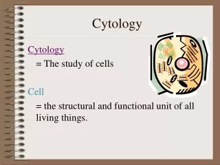
Cytology The key to every biological problem must finally be sought in the cell, for every living organism is, or at some time has been, a cell. E.B. Wilson, 1925
Cell Theory - A good place to start! • Cell Theory was proposed independently by two different individuals in 1838 & 1839 by Schwann & Schleiden. Even though there was collaboration (Schwann animal cell & Schleiden plant cell), Schwann took credit and proposed: 1) The cell is the unit of structure, physiology, and organization in living things. 2) The cell retains a dual existence as a distinct entity and a building block in the construction of organisms. 3) Cells form by free-cell formation, similar to the formation of crystals (spontaneous generation). This last proposal obviously has been proven false! • The modern ideas of the Cell Theory include: 1. all known living things are made up of cells. 2. the cell is structural & functional unit of all living things. 3. all cells come from pre-existing cells by division. 4. cells contains hereditary information which is passed from cell to cell during cell division. 5. All cells are basically the same in chemical composition. 6. all energy flow (metabolism & biochemistry) of life occurs within cells.
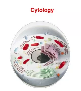
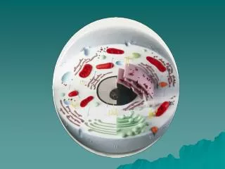
Cell Anatomy • The generalized cell contains: • Cell membrane • Cytoplasm • Cytosol • Organelles • Nucleus
The Generalized Cell – diagram • A few examples…
Cell Membrane Functions • Maintain correct concentration (ratio) of ions between the fluid outside of the cell (extracellular fluid or interstitial fluid) and the fluid inside the cell (intracellular fluid). • Prevent movement of harmful substances in (or at least try to). • Control movement of materials into and out of the cell • Allows for the cell to be sensitive to changes in its environment. • Some membrane features give structural support and integrity to the cell Gatekeeper functions!
The Cell Membrane – The Gate Keeper • The cell membrane has a unique complexity which allows it to act as a selectively permeable membrane. • The basic structural components are: • The phospholipid bilayer membrane • Membrane proteins • Membrane carbohydrates • Cholesterol
The Phospholipid bilayer • The cell membrane is composedof many phospholipid molecules • Consists of a polar “head” and a non-polar “tail” linked by a chargedphosphate molecule (creates the polarity) • This structure allowsthe membrane to“self assemble” intoa lipid bilayer • Hydrophobic tails in • Hydrophilic heads out
The Phospholipid Bilayer & Cholesterol • The phospholipid tails have many cholesterol molecules embedded in them... • This prevents the tails from mass hydrogen bonding which would create a more rigid or less fluid membrane
The Phospholipid Bilayer – Membrane Proteins • Membrane proteins are classified by their location with respect to the membrane. • Through the membrane. . . • These are transmembrane (integral) proteins and have portions exposed to both the intracellular and extracellular fluid • On and in the membrane, but not through. . . • These are peripheral proteins • Membrane protein functions. • Receptor proteins • Channel proteins • Carrier proteins • Enzymes • Anchoring proteins • Cell identification proteins
The Phospholipid Bilayer – Membrane Carbohydrates • Multifunctioning structures often combined with proteins and lipids • Forming glycoproteins & glycolipids • Function in • Cell adhesion • Cell recognition • Cell to cell adhesion • Cell receptor components
The Phospholipid Bilayer – Membrane Transport • The movement of materials between the ECF and ICF is dependent on the cell membrane and it’s components • Movement occurs both with energy input (active) and without energy input (passive) • Below is a link to a pretty good membrane animation site. http://www.wiley.com/legacy/college/boyer/0470003790/animations/membrane_transport/membrane_transport.htm
Passive Transport Across the Membrane • Passive transport requirements: • A gradient • May be pressure, concentration, or any other type of gradient • Movement occurs down a gradient • i.e. from high concentration to lower concentration • Passive transport methods: • Diffusion • Osmosis • Facilitated Diffusion (type of carrier mediated transport) • Filtration
Passive Transport – Diffusion & Osmosis • Diffusion is the movement of molecules other than water from an area of high concentration to an area of low concentration Diffusion Animation • Osmosis is the movement of water from an area of high water concentration to low water concentration. Osmosis Animation
Osmosis & Osmotic Pressure • With a semi-permeable membrane, there will be differences between ECF and ICF composition, these differences will cause water to move to areas of high solute (low water) and in doing so create a pressure (osmotic pressure). • Within a cell, changes in the ECF or ICF composition lead to changes in osmotic pressure in cells) • Isotonic – no net movement of water as osmotic pressure is stable • Cell stays the same • Hypotonic – net movement of water into the cell as the solute level inside the cell is high compared to the outside • Cell expands (may lyse or burst) • Hypertonic – net movement of water out of the cell as the solute level outside the cell is high compared to the inside. • Cell shrinks! (crenates)
Iso-, Hypo-, and Hypertonic Conditions and Cells • http://physioweb.med.uvm.edu/bodyfluids/osmosis.htm
Passive Transport - Filtration • Filtration – created by hydrostatic forces across a membrane • Rate depends on the pressure, the membrane and the solutes present
Passive Transport – Facilitated Diffusion • Use of a carrier molecule in the cell membrane to facilitate the movement of a substance across the membrane • Molecule binds to receptor site on the transmembrane protein • Shape changes due to binding • Molecule is released on the other side!
Active Transport • Requires energy • Occurs against a concentration gradient • Materials can be pumped regardless of gradient direction • Active transport can be: • Carrier mediated transport • Vesicular transport
Carrier Mediated Active Transport • Membrane proteins that are capable of utilizing ATP to change the shape of the transport protein • Movement can be • Single ion (ion pump) in either direction • Multiple ions in two directions as an exchange (counter transport) pump
Non-specific Vesicular Transport • The movement of materials into and out of the cell when wrapped in cell membrane • Can be • Endocytosis – into the cell • Receptor mediated endocytosis - Specific • Pinocytosis – cell drinking • Phagocytosis – cell eating • Exocytosis – out of the cell • Materials ejected may be hormones, waste, mucous, enzymes…
The Cytoplasm • Consists of the cytosol and organelles • Everything inside between the cell membrane and nuclear envelope
The Cytosol • The intracellular fluid (ICF) • Contains nutrients, ions, proteins, wastes & inclusions • Inclusions are often storage units (glycogen & fat) • Contents differ between cells and ECF • Higher [K+] and lower [Na+] in the ICF • More soluble proteins in the ICF – makes the cytosol more viscous, these are involved in cellular metabolic events • ICF contains some carbohydrates (energy), and lots of amino acids (structures) and lipids (energy storage)
production, modification & transport organelles protection & cell regulation organelles energy organelle Organelles • These are the mini functional units of the cell and include • The cytoskeleton • Associated structures of cilia, flagella, centrioles and microvilli • Endoplasmic reticulum • Smooth & Rough • Ribosomes • Proteasomes • Golgi apparatus • Lysosomes • Peroxisomes • Mitochondria
The Cytoskeletal Components • The cytoskeleton is not only a structural support system (framework of the cell), but also enables cellular movement to occur! • Diapedesis • Phagocytosis & exocytosis • Network for motor proteins to “walk” along • Consists of three different filament structures, based on size • Smallest = microfilaments – movement (actin & myosin) • Middle = intermediate filaments – cell adhesion, strength (keratin) • Largest = microtubules – major cytoskeletal component • Cell strength • Rigidity • Anchoring organelles • Creates movement in cilia, flagella and centrioles
The Cytoskeleton in Action A white blood cell using the cytoskeleton to “reach out” for a hapless bacterium.
Centrioles, Cilia & Flagella • More complex structures formed by microtubules • Centrioles – found in animal cells that undergo mitosis • These organelles produce the mitotic spindle that directions chromosomal movement during mitosis • Cilia – a unique arrangement of microtubles in a cylindrical fashion (9+2) • Creates a wave-like coordinated motion for movement of materials along a surface • Flagella • Similar to cilia, but longer and for movement of the cell rather than materials across it
Organelles - Endoplasmic Reticulum • An extensive network of membranes connected to the nuclear envelope • The ER’s job is to • Synthesize • Proteins, hormones, cholesterol, lipids • Storage • Of synthesized materials from the cytosol • Transport • As synthesis occurs, compounds are moved • Detoxification • Enzymes within can destroy toxic compounds
Organelles - Endoplasmic Reticulum • Rough Endoplasmic Reticulum • Network of membranes that contain integrated ribosomal units • Protein production for export • Proteins for enzymes within the ER • Smooth Endoplasmic Reticulum • ER without the ribosomes integrated • Functions to • Make cell membrane phospholipids & cholesterol • Steroid hormone production (estrogens & testosterones) • Synthesis and storage of • Triglycerides and glycogen
Organelles - Ribosomes • Protein manufacturing organelles • Consist of two subunits • May be • Free • proteins made here are used in the cell • Fixed - on endoplasmic reticulum • proteins made here are marked for export from the cell
Organelles - Proteasomes • Opposite actions of ribosomes • Remove proteins by protease (enzymatic) activity • Why? • Removal of damaged or non-functioning proteins
Organelles – Golgi Apparatus • Appears as a set of squished disks on top of each other • Involved with the modification, packaging and transport of ER products • Enzymes to lysosomes • Substances to be secreted (hormones for ex.) • Membrane components
Organelles - Lysosomes • Lysosomes are vesicles filled with digestive enzymes which are activated upon membrane fusion • Destroy phagocytozed material • Removal of damaged organelles • cell death (autolysis & apotosis) • Produced via golgi apparatus…
Organelles - Peroxisomes • Similar to lysosomes, but derived from growth and division of other peroxisomes and contain different active ingredients! • Absorb and break down fatty acids • Produce free radicals (peroxides) as a result, but enzymes convert them into water and oxygen to protect tissues
Organelles - Mitochondria • Often described as the “cell powerhouse” • Functions to • Produce ATP via Kreb’s cycle and the electron transport chain during aerobic conditions (aerobic respiration) • Structurally • Has a double membrane which functions in the above process • Double membrane is folded (cristae) which increases surface area for metabolic reactions to take place • Also contains maternal DNA
The Nucleus • Characteristics: • Typically the most obvious component of a cell (especially under light microscopy) • Contains one or more nucleoli (nucleolus = singular) • Dark/dense areas in the nucleus are sites for synthesis of rRNA and where ribosomal subunits are put together. • Membrane bound (with many nuclear pores) • Control center of the cell • DNA strands contain your entire genetic code in each cell (except mature blood) • Uncondensed DNA = chromatin (not visible) • Condensed = chromosome (visible) • The instructions for all protein synthesis (400,000+ different proteins) • Single nucleus in most cells • Exceptions: skeletal muscle (multiple) & blood (none)
Condensing the DNA to fit (fyi) Higher developed organisms face the problem to store and retrieve a huge amount of genetic information - and this in each cell separately. For instance, the human genome corresponds to 3 billion base pairs (bp) of the DNA double helix, two copies of which make up two meters of DNA chains that have to be stored within the tiny micron-sized nucleus of each cell. These two meters are composed of 46 shorter DNA pieces, each of which, if not condensed, would form a swollen coil of roughly 100 micrometer diameter - clearly much too large to fit into the nucleus. Therefore a suitable compaction mechanism is required. This mechanism, however, should at the same time allow for certain proteins to access specific portions of the DNA and hide (silence) other parts.
Using the genetic Code • DNA contains the “code” for construction of proteins in the pattern of arrangement of nitrogenous base pairs. • Cytosine (C) and Guanine (G) form a base pair as to adenine (A) and thymine (T) • RNA replaces the T with uracil (U)
Using the genetic code • So… we have a section of DNA that we want to make a protein from – how? • Transcription first • Translation second
Transcription • Reading the sequence of base pairs on DNA and making a complementary strand of RNA (called messenger RNA) • Steps: • RNA polymerase binds to promoter region of gene to be transcribed • RNA directs new base pairs as it moves along the codons (three base pair units) • RNA polymerase stops when it reaches a stop codon Detailed Animation of Transcription & Translation
Translation • This process take the mRNA created during transcription and “reads” the codons for directions in assembling amino acids together to form proteins. • Steps: • mRNA binds to small ribosomal subunit at the start codon (AUG) • A tRNA with matching anticodon arrives carrying an amino acid • The ribosomal units continue to “read” the codons and direct adittional tRNA units with matching anti-codons and their corresponding amino acids • A peptide bond (dehydration synthesis) is formed between the amino acids creating a single chain of amino acids – a peptide • The ribosmal units reach the “stop” codon and disengage from the mRNA • This process can be occurring many times simultaneously on one mRNA!
Cell Life Cycle • A cell’s life typically involves a period of division (mitotic phase), and the normal cell processes and growth phases (interphase) • G1 – normal cell functioning along with organelle duplication and cell growth • S – DNA replication and histone synthesis – preparation! • G2 – more normal cell functioning (protein synthesis)
Mitotic Phase • Mitosis – a multi-step process leading to duplication of the cell upon completion of cytokinesis • Steps of Mitosis: • Prophase • Metaphase • Anaphase • telophase
Prophase • Prophase • early in prophase the chromosomes condense and become visible under special stain. • The nuclear membrane breaks down and the spindle apparatus begins to form.
Metaphase • The condensed chromosomes (chromatids) line up at the metaphase plate