CODE: 1Agc00004
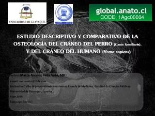
CODE: 1Agc00004. ESTUDIO DESCRIPTIVO Y COMPARATIVO DE LA OSTEOLOGÍA DEL CRÁNEO DEL PERRO ( Canis familiaris ), Y DEL CRÁNEO DEL HUMANO ( Homo sapiens ). Autho r: Marco Antonio Véliz Aubá, MV /

CODE: 1Agc00004
E N D
Presentation Transcript
CODE: 1Agc00004 ESTUDIO DESCRIPTIVO Y COMPARATIVO DE LA OSTEOLOGÍA DEL CRÁNEO DEL PERRO (Canis familiaris), Y DEL CRÁNEO DEL HUMANO (Homo sapiens) Author: Marco Antonio Véliz Aubá, MV / e-mail: marcoveliz@chile.comInstitution: Taller de preparaciones anatómicas. Escuela de Medicina. Facultad de Ciencias Médicas. Universidad de Guayaquil, EcuadorYear: 2009Language: Spanish
ABSTRACT Human being and domestic canine have experienced millions of years of independent evolution since a commom ancestor. By means of the analysis of 3 human skulls (Homo sapiens) and 3 mesaticephalic type canine skulls (Canis familiaris), previously treated by osteological study techniques, structural similarities and differences in quantity and quality terms were compared. COMPARATIVE AND DESCRIPTIVE STUDY OF DOG SKULL (Canis familiaris) AND HUMAN SKULL (Homo sapiens) OSTEOLOGY.
ABSTRACT continuation. Comparison of dentition, form and bone amount of neuro and splachnocranial elements was made. Morphological resemblances were undeniable, demonstrating the homology and proving thereby the existence of common ancestors. The most remarkable differences were found in the orbital cavity so as incisive and interparietal bone belonging exclusively to dog skulls. All these characteristics are directly related to the functional development of each of the studied species. COMPARATIVE AND DESCRIPTIVE STUDY OF DOG SKULL (Canis familiaris) AND HUMAN SKULL (Homo sapiens) OSTEOLOGY.
ESTUDIO DESCRIPTIVO Y COMPARATIVO DE LA OSTEOLOGÍA DEL CRÁNEO DEL PERRO (Canis familiaris) Y DEL CRÁNEO DEL HUMANO (Homo sapiens) RESUMEN El ser humano y el canino doméstico han sufrido millones de años de evolución independiente a partir de un ancestro común. Mediante el análisis de 3 cráneos humanos (Homo sapiens) y 3 cráneos de perro (Canis familiaris) tipo mesocefálico previamente tratados bajo métodos de Osteotecnia, se compararon estructuralmente similitudes y diferencias en términos cualitativos y cuantitativos. Se comparó dentición, forma y número óseo de Neuro y esplácnocraneo.
ESTUDIO DESCRIPTIVO Y COMPARATIVO DE LA OSTEOLOGÍA DEL CRÁNEO DEL PERRO (Canis familiaris) Y DEL CRÁNEO DEL HUMANO (Homo sapiens) RESUMEN continuación Las semejanzas morfológicas fueron evidentes, demostrando la homología y comprobando así la existencia de antepasados en común. Las diferencias más marcadas se encontraron en la cavidad orbitaria, y en huesos de exclusiva presencia en el canino, tales como el hueso incisivo y el interparietal. Todas estas características directamente relacionadas con el desarrollo funcional de cada una de las especies estudiadas.
Clasificación Hombre Reino: Animalia Phylum: Chordata Subphylum: Vertebrata Clase: Mammalia Orden: Primates Familia: Hominidae Género: Homo Especie: H. Sapiens Clasificación Perro Reino: Animalia Phylum: Chordata Subphylum: Vertebrata Clase: Mammalia Orden: Carnivora Familia: Canidae Género: Canis Especie: C. lupus Subespecie: C. familiaris
HUESOS NEUROCRANEO HUESOS ESPLACNOCRANEO
Figura 3.- Mandíbula. Canisfamiliaris 1.- Proceso coronoideo 2.- Cresta coronoidea 3.- Fosa masetérica 4.-Agujero mentoniano 5.- Proceso angular 6.- Proceso condilar 7.- Línea milohioidea 8.- Primer molar 9.- Rama mandibular 10.- Cuerpo mandibular 11.- Sínfisis mentoniana 13.- Agujero mandibular 14.- Cresta condilar Figura 2.-. Cráneo Canis familiaris, vista caudal.
Figura 6.-. Cráneo Canis familiaris, vista ventral. 1.- Hueso incisivo 2.- Dientecanino 3.- Fisura palatina 4.-Surco palatino 5.- Cuarto premolar 6.- Agujero palatino mayor 7.- Lamina horizontal del palatino 8.- Hueso Pterigoideo 9.- Hueso preesfenoides 10.- Hueso basiesfenoides 11.- Agujero oval 12.- Agujero carotideo externo/ 13.- Bulla timpánica 14.- Fisura timpanoccipital 15.- Proceso yugular 16.- Proceso condilar (cóndilo occipital) 17.- Agujero magno 18.- Canal hipogloso 19.- Espina nasal caudal del palatino 20.- Tubérculo faríngeo Flechas.-Coanas
Figura 7.-. Cráneo canino y cráneo humano, vista lateral. Na.- Hueso Nasal P.- Hueso Parietal C.- Hueso Cigomático T.- P. escamosa del Temporal F.- Hueso Frontal O.- Hueso Occipital Pt.- Pterigoides M.- Hueso Maxilar L.- Hueso Lagrimal In.- Hueso Incisivo
Figura 8.1.-. Cráneo canino y cráneo humano, vista rostral. Na.- Hueso Nasal CN.- Cavidad nasal AI.- Agujero infraorbitario F.- Hueso Frontal L.- Hueso Lagrimal M.- Hueso Maxilar N.- Nasion PR.- Prostion In.- Hueso Incisivo
Figura 8.2-. Cráneo canino y cráneo humano, vista dorsal. Na.- Hueso Nasal P.- Hueso Parietal C.- Arco Cigomático T.- Hueso Temporal F.- Hueso Frontal I.- Inion B.- Bregma M.- Hueso Maxilar N.- Nasion PR.- Prostion
Figura 9.-. Vista Craneal. CAVIDAD ORBITARIA 1.- Hueso frontal 2.- Hueso maxilar 3.- Canal óptico 4a.-Fisura orbitaria 4b.-Fisura orbitaria superior 5.- Fisura orbitaria inferior 6.- Hueso cigomático 7.- Proceso cigomático del frontal 8.- Proceso frontal del cigomático 9.- Proceso orbitario lateral del frontal 10.- Proceso frontal del cigomático 11.- Proceso cigomático del temporal
BIBLIOGRAFIA ADAMS, D. (1988). Anatomía canina, Estudio sistémico. Facultad de Veterinaria, Iowa State University. Editorial Acribia. 518p COELLO Cuntó, Rafael. (2004).Fundamentos de Anatomía Humana. Guayaquil. 1a Edición.. 410p COHEN, Meyer . (2006). Michael.Perspectives on the face. Oxford University Press, Inc. New York. CONCHA, Ismael. (1998). Syllabus Anatomía General. Medicina Veterinaria: Capitulo D, Recopilación sobre: Cabeza del canino. Santiago, Chile. EVANS, (1993) H.E. y DELAHUNTA, A. Miller´s Guide to the Dissection of the Dog. 3era Edición. W.B. Saunders Co. Philadelphia. SISSONS, S y GROSSMAN, J.D. (1982) Anatomía de los animales domésticos. 5ta Edición. Tomo I. TESTUD, L y LATARJET, M. (1983). Tratado de Anatomía Humana. Salvat Editores. Madrid. 4 Tomos. ESTUDIO DESCRIPTIVO Y COMPARATIVO DE LA OSTEOLOGÍA DEL CRÁNEO DEL PERRO (Canis familiaris) Y DEL CRÁNEO DEL HUMANO (Homo sapiens)