Download
1 / 63
630 likes | 1.24k Views
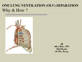
ONE LUNG VENTILATION (OLV)-SEPARATION Why & How ?. By Ahmed Ibrahim ; M.D. Prof.of Anaesthesia Ain Shams University. OLV means: separation of the two lungs each lung functions independently by preparation of the airway OLV provides:

E N D
ONE LUNG VENTILATION (OLV)-SEPARATION Why & How ? By Ahmed Ibrahim ; M.D. Prof.of Anaesthesia Ain Shams University
OLV means: • separation of the two lungs • each lung functions independently by preparation of the airway • OLV provides: • protection of healthy lung from infected/bleeding one • diversion of ventilation away from damaged airway or lung • improved exposure of surgical field • OLV causes: • more manipulation of airway, more damage • significant physiologic change & easy development of hpoxaemia
Indications for OLV ABSOLUTE 1. Isolation of one lung from the other to avoid spillage or contamination. 2. Control of the distribution of ventilation (fistula, cyst, T.B disruption…). 3. Unilateral bronchopulmonary lavage. RELATIVE 1. Surgical exposure. 2. Postcardiopulmonary bypass status, after removal of totally occluding chronic unilateral pulmonary emboli.
OLVis achieved by either; -Double lumen ETT (DLT) -Bronchial blocker -Endobronchial tube
Features of DLT RUL, right upper lobe; LUL, left upper lobe
Carlens DLT Robertshaw DLT
Rt Lt
guide for Length and Size of DLT Length of tube , For 170 cm height, tube depth of 29 cm For every 10 cm height change , 1 cm depth change
Check Position of Lt -DLT Checklist for tracheal placement a. inflate tracheal cuff b. ventilate rapidly by hand c. check that both lungs are being ventilated d. If not, withdraw 2-3 cm & repeat Checklist for Lt side a. inflate Lt cuff > 2ml b. ventilate and check bilateral breath sounds c. clamp Rt tube d. check unilateral (Lt) breath sounds Checklist for Rt side a. clamp Lt tube b. check unilateral (Rt) breath sounds
Major Malpositions of a Lt- DLT Lt Breath Sounds Heard Left Right Both Left None / Very minimal left Both None / Very minimal Both Right None / Very minimal Right
To ensure correct position of DLT clinically : • breath sounds are - normal (not diminished) & - follow the expected unilateral pattern with unilateral clamping • the chest rises and falls in accordance with the breath sounds • the ventilated lung feels reasonably compliant • no leaks are present • respiratory gas moisture appears and disappears with each tidal ventilation N.Beven if the DLT is thought to be properly positioned by clinical signs, subsequent FOB may reveal an incidence of malposition ( 38 -78 %)
Other Methods to Check DLT Position • Chest radiograph ; may be more useful than conventional auscultation and clamping in some patients, but it is always less precise than FOB. The DLT must have radiopaque markers at the end of Rt and Lt lumina. • Comparison of capnography; waveform and ETCO2 from each lumen may reveal a marked discrepancy (different degree of ventilation). • Surgeon ; may be able to palpate, redirect or assist in changing DLT position from within the chest (by deflecting the DLT away from the wrong lung, etc..).
Complications of DLT • impediment to arterial oxygenation for OLV • tracheobronchial tree disruption, due to -excessive volume and pressure in bronchial balloon -inappropriate tube size -malposition • traumatic laryngitis (hook) • inadvertent suturing of the DLT
to avoid Tracheobronchial tree Disruption ; 1. Be cautious in patients with bronchial wall abnormalities. 2. Pick an appropriately sized tube. 3. Be sure that tube is not malpositioned ; Use FOB. 4. Avoid overinflation of endobronchial cuff. 5. Deflate endobronchial cuff during turning. 6. Inflate endobronchial cuff slowly. 7. Inflate endobronchial cuff with inspired gases. 8. Do not allow tube to move during turning.
Relative Contraindications to Use of DLT • full stomach (risk of aspiration); • lesion (stricture, tumor) along pathway of DLT (may be traumatized); • small patients; • anticipated difficult intubation; • extremely critically ill patients who have a single-lumen tube already in place and who will not tolerate being taken off mechanical ventilation and PEEP even for a short time; • patients having some combination of these problems. Under these circumstances, it is still possible to separate the lungs by : -using a single-lumen tube + FOB placement of a bronchial blocker ; or -FOB placement of a single-lumen tube in a main stem bronchus.
Bronchial Blockers(With Single-Lumen Endotracheal Tubes) • Lung separation can be effectively achieved with the use of a single-lumen endotracheal tube and a FOB placed bronchial blocker. • Often necessary in children as DLTs are too large to be used in them. The smallest DLT available is a left-sided 26 Fr tube, which may be used in patients 8 -12 years old and weighing 25 -35 kg. • Balloon-tipped luminal catheters have the advantage of allowing suctioning and injection of oxygen down the central lumen.
Indications for Use of Bronchial Blockers 1st , limitations to DLT (severely distorted airway, small patients , anticipated difficult intubation) 2nd, to avoid a risky change of DLT to single-lumen tube • whenever postoperative ventilation is anticipated • in cases of thoracic spine surgery in which a thoracotomy in the supine or LDP is followed by surgery in the prone position. 3rd , situations in which both lungs may need to be blocked (e.g., bilateral operations, indecisive surgeons).
Types of bronchial blockers • Univent bronchial blocker system • Arndt endobronchial blocker • Cohen Flexitip Endobronchial Blocker • BB independent of a single-lumen tube
steps of FOB-aided method of positioning the Univent bronchial blocker in lt main stem bronchus One- or two-lung ventilation is achieved simply by inflating or deflating, respectively, the bronchial blocker balloon
Advantages of the Univent Bronchial Blocker Tube ( Relative to DLT ) 1. Easier to insert and properly position. 2. Can be properly positioned during continuous ventilation and in the lateral decubitus position. 3. No need to change the tube when turning from the supine to prone position or for postoperative mechanical ventilation. 4. Selective blockade of some lobes of each lung. 5. Possible to apply CPAP to nonventilated operative lung.
Arndt endobronchial blocker[Wire guided Endobronchial Blocker (WEB)]
Bronchial Blockers that are Independent of a Single-Lumen Tube Adults -Fogarty (embolectomy) catheter with a 3 ml balloon. It includes a stylet so that it is possible to place a curvature at the distal tip to facilitate entry into the larynx and either mainstem bronchus . -balloon-tipped luminal catheters (such as Foley type) may be used as bronchial blockers. Very small children (10 kg or less) - Fogarty catheter with a 0.5 ml balloon - Swan-Ganz catheter (1 ml balloon) * these catheters have to be positioned under direct vision; a FOB method is perfectly acceptable; the FOB outside diameter must be approximately 2 mm to fit inside the endotracheal tube (3 mm internal diameter or greater). Otherwise, the bronchial blocker must be situated with a rigid bronchoscope. * Paediatric patients of intermediate size require intermediate size occlusion catheters and judgment on the mode of placement (i.e., via rigid versus FOB).
Lung separation with a single-lumen tube, FOB, and Rt lung bronchial blocker
Disadvantages of a blocker that is independent of the single-lumen tube as compared with DLT • inability to suction and/or to ventilate the lung distal to the blocker. • increased placement time. • the definite need for a fiberoptic or rigid bronchoscope. • if bronchial blocker backs out into the trachea, the seal between the two lungs will be lost and the trachea will be at least partially obstructed by the blocker, and ventilation will be greatly impaired.
Endobronchial Intubation with Single-Lumen Tubes • In adults, is often the easiest, quickest way for lung separation in patients presenting with haemoptysis , either -blind, or -FOB , or -guidance by surgeon from within chest • In children it may be the simplest way to achieve OLV Disadvantages -inability to do suctioning or ventilation of operative side. -difficult positioning bronchial cuff with inadequate ventilation of Rt upper lobe after Rt endobronchial intubation.
In summary, DLT is the method of choice for lung separation in most adult patients. The precise location can be determined by FOB . In situations where insertion of a DLT may be difficult and/or dangerous, separating the lungs is achieved either with a single-lumen tube alone or in combination with a bronchial blocker (e.g., the Univent tube). Therefore, regardless of what method of lung separation chosen, there is a real need of a small-diameter FOB (for checking the position of the DLT, placing a single-lumen tube in a mainstem bronchus, and placing a bronchial blocker) .
Physiology of OLV (Arterial Oxygenation and Carbon Dioxide Elimination) Blood passing through : • non ventilated lung ,retains CO2 and does not take O2. • over ventilated lung ,gives off more than a normal amount of CO2 but cannot take up a proportionately increased amount of O2 .
Thus, during one-lung ventilation • more decreased oxygenationthan during two-lung ventilation in LDP due to an obligatory Rt-Lt transpulmonary shunt through the nonventilated nondependent lung. Consequently, lower PaO2 & larger P(A-a)O2 • usually carbon dioxide elimination is not a problem; but retention of CO2 by blood traversing the nonventilated lung slightly exceeds the increased elimination of CO2 from blood traversing the ventilated lung, and the PaCO2 will usually slowly increase and P(A-a)CO2 decreases .
Two-lung ventilation versus OLV during OLV, the nonventilated lung has some blood flow and therefore has an obligatory shunt, which is not present during two-lung ventilation & is the most important reason for increased P(A-a)O2.
Blood Flow distribution during OLV • The major determinants of blood flow distribution between both lungs : • gravity, • amount of lung disease, • magnitude HPV, • surgical interference nondependent , • ventilation mode dependent
Blood Flow Distribution During OLV , cont…. • Lung condition (amount of lung disease) *severely diseasednondependent lung, may have a fixed reduction in blood flow preoperatively and its collapse may not cause much increase in shunt. *increases in PVR in dependent ventilated lung decreases its ability to accept redistributed blood from the hypoxic lung. This may occur in case of : -decreasing FIO2 in the dependent lung . -decreasing temperature .
Blood Flow Distribution During OLV , cont…. • Also, development of a hypoxic compartment (area of low V/Q and atelectasis) in the dependent lung increases its PVR (HPV), thereby decreasing dependent lung and increasing nondependent lung blood flow. • This may develop intraoperatively for several reasons: • 1. in LDP ,ventilated dependent lung usually has • a reduced volume resulting from combined factors • of induction of anaesthesia and circumferential • compression by mediastinum ,abdominal contents, and • suboptimal positioning effects (rolls, packs, supports). • 2. absorption atelectasis can occur in regions with low V/Q when they are exposed to high FIO2 . • 3. difficulty in secretion removal . • 4.maintaining the LDP for prolonged periods may cause fluid to transude into the dependent lung and cause further decrease in lung volume and increase in airwayclosure.
Blood Flow Distribution During OLV , cont. • Surgical interference(compression ,retraction and ligation of pulmonary vessels during pulmonary resection) of the nondependent lung may further passively reduce its blood flow. • Mode of ventilation of dependent lung • If hyperventilated PaCO2 HPV • Excessive AWP (PEEP or VT ) dependent PVR and nondependent lung blood flow. • FIO2 -VD in dependent lung, augmenting HPV in nondependent lung -but ,may cause absorption atelectasis in regions that have low V/Q ratios
Blood Flow Distribution During OLV , cont. • Magnitude of HPV • HPV is an autoregulatory mechanism that protects the PaO2 by decreasing the amount of shunt flow that can occur through hypoxic lung as it diverts blood flow from the atelectatic lung toward the remaining normoxic or hyperoxic ventilated lung. • HPV is of little importance When ; -very little of the lung is hypoxic (near 0%) because shunt will be small. -most of the lung is hypoxic (near 100%) there is no significant normoxic region to which the hypoxic region can divert flow. • Of great importance if the percentage of hypoxic lung is intermediate ( 30 and 70%), which is the case during OLV
Blood Flow Distribution During OLV , cont. Factors that might determine the amount of regional HPV