Download
1 / 26
400 likes | 1.1k Views
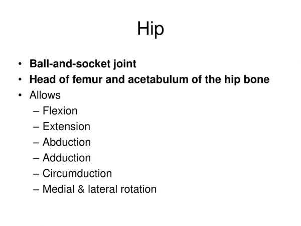
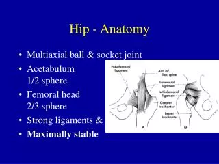
Hip - Anatomy. Multiaxial ball & socket joint Acetabulum 1/2 sphere Femoral head 2/3 sphere Strong ligaments & capsule Maximally stable. Anatomy. Forces Standing - 0.3 times body weight Standing on 1 leg - 2.5 times body weight Walking - 1.3 to 5.8 times body weight

E N D
Hip - Anatomy • Multiaxial ball & socket joint • Acetabulum1/2 sphere • Femoral head2/3 sphere • Strong ligaments & capsule • Maximally stable
Anatomy • Forces • Standing - 0.3 times body weight • Standing on 1 leg - 2.5 times body weight • Walking - 1.3 to 5.8 times body weight • Walking up stairs - 3 times body weight • Running - 4.5+ times body weight
History • Age • infancy: congenital hip dysplasia • 3-12 year old boys: Legg-Calve-Perthes Dz • middle age & elderly: osteoarthritis • Mechanism of injury • land on outside hip • land on knee • repetitive loading
History • Pain details • location • snapping • progression of symptoms • exacerbating factors • alleviating factors • Weakness • Occupation, Sport
Observation • Gait • Posture • Balance • Limb position • shortened, adducted, medially rotated • abducted, laterally rotated • shortened, laterally rotated • Leg shortening
Examination • Active Range of Motion • Flexion: 110 to 120 degrees • Extension: 10 to 15 degrees
Examination • Active Range of Motion • Abduction: 30 to 50 degrees • Adduction: 30 degrees
Examination • Active Range of Motion • External rotation: 40 to 60 degrees • Internal rotation: 30 to 40 degrees
Examination • Passive Range of Motion • Intra-abdominal inflammation may cause pain with passive medial & lateral hip rotation • Hip pathology indicated by groin discomfort and decreased ROM on medial rotation • Acetabular rim or labral problems with click and painful hip flexion, abduction, andmedial rotation
Examination • Strength testing • isometric • eccentric • knee extension • knee flexion
Examination • Functional Testing • squatting • up & down stairs one at a time • crossing legs • up & down stairs two at a time • running straight ahead • running and decelerating • running and twisting • one-legged hop
Special Tests • Patrick’s Test(Faber, Figure 4) • supine • foot on opposite knee • hip involvementiliopsoas spasmSI joint involvement
Special Tests • Ortolani’s & Barlow’s • Assess for congenital dislocation of hips • Valid for first 6 monthsof life • Positive test is “clunck” not “click”
Special Tests • Galeazzi Test • knees & hips flexed to 90 degrees • positive test: one knee higher • Telescoping Sign • knee & hip flexed to 90 degrees, axial load and distraction applied • positive test: increased relative movement
Special Tests • Leg length • true leg length discrepancycongenital maldevelopmenttrauma • functional leg length discrepancyscoliosis
Special Tests • Leg length • Measured from ASIS to medial malleolus • Functionally measured • knees & hips flexed with thumbs on medial malleolus then knees and hips extended
Special Tests • Flexibility • modified Thomas Test • assesses both hip flexor and quad flexibility
Special Tests • Flexibility • Ober’s Test • assesses iliotibial band flexibility
Special Tests • Flexibility • Piriformis Test • Assessess flexibility of piriformis muscle • “Piriformis Syndrome”
Special Tests • Flexibility • Popliteal Angle • Assesses hamstring flexibility
Palpation • Iliac crest Greater trochanter • ASIS Ischial tuberosity • PSIS
Diagnostic Imaging • Radiographs • Anterior-Posterior view • Frog leg view • CT • MRI • Arthrogram
Case Studies • 7 year old boy with 5 week h/o limp • Limp is more pronounced when he’s tired • Complains of left knee pain
Case Studies • 55 year old male with right hip & back pain • He notes sciatica and groin pain • Symptoms worsen with walking • Very active lifestyle
Case Studies • 18 year old female hurt her hip surfing • Hip is medially rotated and shortened • She notes sciatic pain
Case Studies • 12 year old large boy with limp • Fell from chair onto floor and complains of right knee and thigh pain