Understand the Absorption Process in the Digestive System
270 likes | 304 Views
Learn about the absorption of digested food in the gastrointestinal tract, particularly in the ileum of the small intestine. Discover the structures and mechanisms involved in the absorption process and the adaptations of the ileum for efficient absorption of nutrients.

Understand the Absorption Process in the Digestive System
E N D
Presentation Transcript
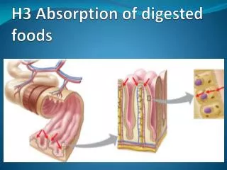
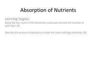
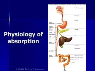
Absorption of Digested Food Absorption is the uptake of digested food molecules from the alimentary canal (gastrointestinal tract) into the blood or lymph The principal site for absorption of nutrients is the ileum of the smallintestine where the epithelialcells lining this gut region are ideally suited to this role Glucose and aminoacids are transported across the epithelial cells to the bloodcapillaries within the villi, and reconstituted fats are absorbed into the lymphcapillaries (lacteals)
The gross and histological structure of the ileum adapt this region for its absorptive and secretory roles Absorption of Digested Food Absorption involves the transportprocesses of simple diffusion, facilitated diffusion, active transport and osmosis (for water absorption)
gland within the submucosa (Brunner’s glands) gland outside the gut, e.g. pancreas mucosa villi muscularis mucosa submucosa (connective tissue) circular muscle longitudinal muscle outer serosa lumen
The duodenum is the site where pancreaticjuice (containing many enzymes) is secreted into the gut and where Brunner’sglands secrete an alkalinemucus to help neutralise and protect its lining from the acidchyme arriving from the stomach The ileum is the principal site for the absorption of nutrients; it is verylong (about 4 metres in length) and the numerous villi with their epitheliallinings increase the surfacearea for absorption
Adaptations of the Ileum The ileum displays adaptations for both absorption and secretion: • The ileum is very long and absorption can occuralong its length • The mucosa is highlyfolded and the numerous ‘finger-like’ projections, the villi, vastly increase the surfacearea of the epithelium for both digestion and secretion • The epithelial cells of the villi bear microvilli at their luminal surface that project into the lumen of the gut; these microvilli form the brushborder and further increase the surface area available for both absorption and secretion • The villi are well supplied with a networkofbloodcapillaries into which glucose and amino acids are transferred and then transported to the liver along the hepatic portal vein • A single, permeable lacteal within each villus transports reconstituted fats away from the intestine
Epithelial cells, bearing microvilli, project into the lumen of the gut Intestinal gland between the villi; contains enzyme secreting cells and hormone releasing cells
T.S. Ileum intestinal gland Lumen villi mucosa
Microvilli forming the brush border Numerous mitochondria Nucleus Microvilli further increase the surface area of the intestine for efficient absorption of food molecules Numerous mitochondria provide energy, in the form of ATP, for the active transport of various molecules and ions
mucus-secreting, goblet cell in the epithelial lining of the villus epithelial cells brush border
brush border goblet cell epithelial cell nucleus of epithelial cell
microvilli of epithelial cells (brush border) mitochondrion
Brush border Digested food molecules are transported across the epithelial cells of the villi into the blood capillaries (monosaccharides and amino acids) and the lacteals (fatty acids and monoglycerides) Interior of epithelial cells
Mucosal membrane Serosal membrane Brush border Each epithelial cell has a mucosal and a serosal membrane The mucosal membrane of the microvilli projects into the lumen of the gut with the serosalmembrane being closely apposed to the blood capillaries within the villus Interior of epithelial cells
Membrane-bound protein carriers are involved in the transport of glucose molecules by facilitated diffusion and active transport Glucose is transported across the epithelialcells of the villi from the lumen of the gut to the blood capillaries by facilitateddiffusionand sodium-linked activetransport Facilitated diffusion is dependent upon the existence of a glucoseconcentrationgradient across the epithelial cell membranes Active transport allows for the transfer of glucose against its concentration gradient
Facilitated diffusion is a carrier-assisted transport mechanism in which molecules are transferred across membranes along their concentration gradients
Glucose is transported into the epithelial cells of the villi by facilitated diffusion as long as a concentration gradient exists between the gut lumen and the interior of the cells
Active transport is a carrier-assisted, energy requiring transport mechanism that utilises energy from ATP molecules to transfer molecules against their concentration gradient
Various membrane-bound carrierproteins in the epithelial cells of the villi are involved in the sodium-linked, activetransport of glucose from the gut lumen to the blood capillaries Specific carriers located within the mucosal membrane possess binding sites for both glucose and sodium ions This co-transport of glucose and sodium ions into the cell is dependent upon a sodium ion concentration gradient
Specific protein carriers in the mucosal membranes of intestinal epithelial cells possess binding sites for both glucose molecules and sodium ions The transport of these molecules into the epithelial cells from the gut lumen is dependent upon the sodium ion concentration gradient; so long as this gradient exists, glucose can be transported against its own gradient
The sodium gradient is maintained by sodium/potassium pumps located within the serosal membrane of the epithelial cells These energy-requiring pumps transfer sodium ions out of the cell into the blood in exchange for potassium ions The operation of these Na+/K+ pumps results in a permanently low Na+ concentration within the epithelial cells and the maintenance of a Na+ concentration gradient across the mucosal membrane
Once inside the epithelial cells, glucose molecules are transferred across the serosal membranes into the blood capillaries by facilitated diffusion In this system, the transport of glucose across the mucosal membranes is coupled to the active transport of sodium ions across the serosal membranes
Absorption of Amino Acids The mechanism for absorption of amino acids is similar to that for glucose absorption, i.e.sodium-linked active transport The difference between the two processes is such that there are many different co-transportcarriers in the mucosal membrane There are many different amino acids thatdiffer in structure and shape Individual carriers for the co-transport of sodium ions and each amino acid are located within the mucosal membrane
Fatty acids, monoglycerides and glycerol enter the epithelial cells by simplediffusion where they are resynthesised into triglyceride micelles release fatty acids, monoglycerides and glycerol