Download
1 / 41
420 likes | 586 Views
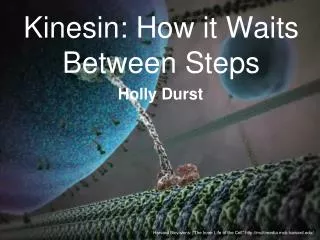
Kinesin: How it Waits Between Steps. Holly Durst. Harvard Biovisions: “The Inner Life of the Cell” http://multimedia.mcb.harvard.edu/. What is Kinesin?. dimeric motor protein carries cellular cargo along microtubules by hydrolyzing ATP

E N D
Kinesin: How it Waits Between Steps Holly Durst Harvard Biovisions: “The Inner Life of the Cell” http://multimedia.mcb.harvard.edu/
What is Kinesin? • dimeric motor protein • carries cellular cargo along microtubules by hydrolyzing ATP • takes several hundred “steps” along a microtubule without detaching Harvard Biovisions: “The Inner Life of the Cell” http://multimedia.mcb.harvard.edu/
Coiled coil β α Neck linkers • ATP binding to leading head initiates neck linker docking and the other head is thrown forward • New leading head docks onto binding site after diffusional search, resulting in 80 Å movement of attached cargo • This accelerates ADP release and trailing head hydrolyzes ATP to ADP-Pi • ATP binds to leading head
FRET http://bio.physics.uiuc.edu/images/FRET_concept.jpg
How kinesin waits between steps Teppei Mori, Ronald D. Vale & Michio Tomishige Objective • Use a series of smFRET experiments to detect whether kinesin is bound to its microtubule track by one or two heads in its ‘waiting’ conformation between steps
The Players • ‘cysteine light’ human ubiquitous kinesin-1 dimer which cysteine residues and/or mutations were introduced into • Dye-labelled kinesins were imaged moving along sea-urchin axonemes with a custom-built prism-type laser-illuminated total-internal reflection fluorescence microscope
Heterodimer one chain containing single cysteine residue in plus end oriented tip of core (residue 215) one chain containing single cysteine residue in minus-end oriented base of the core (residue 43) The Players Used two FRET Sensors:
The Players 2. Homodimer with a cysteine residue in both chains at the beginning of the neck linker (residue 324)
Testing the Sensors • Donor dye: Maleimide modified Cy3 • Acceptor dye: Maleimide modified Cy5 • Molecules that contained both dyes were selected for smFRET observations
Examined FRET efficiency in presence of non-hydrolyzable nucleotide analog AMP-PNP so that both kinesin heads are bound statically to the microtubule Testing the Sensors
Bimodal distribution of low (10%) and high (90%) As expected if two kinesin heads are bound to adjacent tubulin subunits 8 nm apart High peak – 43 dye on leading head and 215 dye on trailing head Low peak – 215 dye on leading head and 43 dye on trailing head Unimodal distribution centered at about 35% Is consistent with a two-head bound state Testing the Sensors smFRET Efficiencies for 215-43 smFRET Efficiencies for 324-324 Binding along single protofilament supported by experiments with a 149-324 sensor
Different Conditions • Low ADP concentrations Remember: ADP occupying binding site = weak microtubule binding 215-43 Sensor unimodal at about 30% • 324-324 Sensor • shift from 35% to 60% • kinesin heads come closer together Under these conditions, these distributions reflect a one-head bound state
Different Conditions • Low ADP plus excess inorganic phosphate • Partial occupancy of an ADP·Pi state in tethered head 324-324 Sensor 215-43 Sensor • Peaks characteristic of a two-head bound state • Similar results for addition of AlF4-
Different Conditions • Both heads nucleotide free 215-43 Sensor 324-324 Sensor • distributions similar to AMP-PNP, but with broader distributions • Nucleotide-free kinesin primarily adopts a two-head bound state with partial occupancy of a one-head-bound state
FRET efficiency trace of individual axoneme-bound 215-43 heterodimer kinesin
Signal of 215-43 in AMP-PNP was fairly constant • A subset of molecules with ADP or ADP/Pi or under nucleotide-free conditions underwent abrupt FRET transitions • Unbinding and rebinding of kinesin head with microtubule
Mutations • Mutated so only one head could bind to microtubules under all conditions • Y274A/R278A/K281A in loop 12 (L12-triple) • 215(WT) – 43 (L12) • 215(L12) – 43 (WT)
Mutations • 200 nM ADP • 215(WT)-43(L12) and 215(L12)-43(WT) both produced unimodal distributions centered at about 30% • Distances between 43-labelled and 215 labelled dyes are similar • Similar result for nucleotide-free state
Mutations • Addition of AMP-PNP • 215(WT)-43(L12) – bimodal with primary peak at 80% • 215(L12)-43(WT) – major peak shifted in opposite direction toward lower efficiencies • movement of L12 triple towards plus-end oriented tip of bound head
Mutations • 215/342 dyes on wild-type chain to probe neck linker conformations in the bound head
Mutations Translation of unbound head from rear position to forward position is driven by nucleotide-dependent docking of neck-linker
Dynamic Measurements • Saturating ATP concentration (1 mM) • Can only measure an average
Dynamic Measurements • 215-43 showed broad distribution centered at about 50% • Average of bimodal 10%, 90% FRET distribution of static two-head bound kinesin • Different from 30% value of one-head bound kinesin
Dynamic Measurements • 324-324 unimodal distribution centered at about 30% Kinesin spends most of the time bound with two heads to the microtubule when moving at saturating ATP concentrations
Dynamic Measurements • Subsaturating ATP concentration (2 μM)
Dynamic Measurements • 215-43 shifted to about 30% • 324-324 shifted to about 60% • More similar to 200 nM ADP (one-head bound) Suggests that kinesin waits primarily as a one-head bound intermediate when ATP binding becomes the rate-limiting step in the ATPase cycle
Dynamic Measurements • Longer dwell times at low ATP concentration
Dynamic Measurements • Spent most time in a roughly 30% FRET state (one-head bound) with brief spikes towards higher (80%) FRET values • Higher values represent transient two-head bound intermediate state • Transitions from 30% to lower FRET state should also occur • Difficult to distinguish from noise
Dynamic Measurements • Dwell-time histogram best fitted by a convolution of two exponentials • Two rate-limiting ATP binding events occur between the two high-FRET spikes
Dynamic Measurements • Mean dwell time (140 ms) is comparable to predicted dwell time • Total number of spikes divided by displacement of these molecules yielded an average distance of about 17 nm per spike • Close to double kinesin step size
Dynamic Measurements • A kinesin step at low ATP concentrations involves a short-lived, two-head bound state, which then undergoes a transition to a longer-lived, one-head bound state
Summary • At high ATP concentration (the rate-limiting step is the detachment of the trailing head triggered by ATP hydrolysis/phosphate release), kinesin moves quickly from one two-head bound state to the next • At low ATP concentration (ATP binding to the leading head is rate-limiting), the trailing head releases its Pi and detaches from the microtubule, producing a long-lived one-head bound state
Discussion • Kinesin waits as either a one-head bound or two-head bound intermediate, depending on ATP concentration and the rate-limiting step
Discussion • ATPase cycles in the two kinesin heads are coordinated during processive motion • Gating model proposes that detached head waits in front of bound head and is in a conformation that prevents it from binding tubulin • But, transient interactions with the microtubule are seen • Additional mechanism must keep detached head from progressing through ATPase cycle until its partner binds ATP
Discussion • Detached head will not release ADP when it is interacting with rear tubulin-binding site • ADP release could occur after the bound head binds ATP and docks the neck-linker, translating the detached head to a forward tubulin-binding site • Results are supported by Guydosh and Block who showed that nucleotide dissociation occurs only when a head is in the forward position • Position dependence controlled by conformation of neck-linker
Future Work • How does the conformation of the neck-linker affect transitions in the ATPase cycle?
References • Mori, T.; Vale, R. D.; Tomishige, M. Nature2007, 450, 750-754 • Vale, R. D.; Milligan, R. A. Science2000, 288, 88-95