Download
1 / 36
490 likes | 939 Views
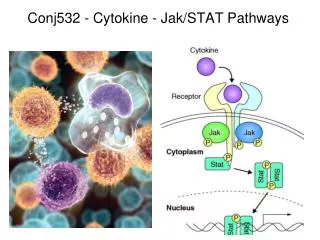
Conj532 - Cytokine - Jak /STAT Pathways. T-helper cell subsets and cytokine profiles .

E N D
T-helper cell subsets and cytokine profiles Th1, Th2 and Th17 cells are a separate lineage of CD4+ T cells, distinct from other T cell subsets. Every specific T helper cells produce its specific cytokines. T-bet, T-box expressed in T cells; FoxP3, forkhead box P3; ROR, retinoid-related orphan receptor. Makoto Kudo, et al. Front Microbiol. 2013;4:263.
Cytokines secreted by immune cells “instruct” T-cells Antigen TH1 like response TH2 like response Effector B cells ('Be1' and 'Be2' cells) can secrete cytokines, such as interferon-γ (IFNγ), interleukin-12 (IL-12), IL-4 and IL-2, that reinforce and stabilize the cytokine profile of effector T helper 1 (TH1) and TH2 cells. In addition, the effector B cells can recruit additional naive T cells into the inflammatory response. Nature Reviews Immunology 10, 236-247
Cellular Mechanisms in Rheumatoid Arthritis Pathogenesis of RA: synovial and systemic inflammation. Inflammation in RA is caused by activation of T cells, B cells and macrophages, which releases cytokines such as IL-1, IL-6 and TNF. These cytokines cause local joint damage through increased production of metalloproteinases and activation of osteoclasts. IL-1, IL-6 and TNF also leak out to the blood stream resulting in systemic inflammation: anaemia, thrombocytosis, fatigue, osteoporosis and the acute-phase response. Abbreviations: IL, interleukin; RA, rheumatoid arthritis. Choy, E. H. et al. Nat. Rev. Rheumatol. 9, 154–163 (2013)
Class I a a a a b (gp130) g b Single chain (dimer) Unique + Common chains (tetramer) Multiple unique chains (2 or 3) IL-2, Interferons IL-6, LIF, IL-11, CNTF), CT-1, CLC, OSM, IL-27 and IL-31. Epo, GH, Prl, GM-CSF 3 General types of cytokine receptors Class II Class III Q: why did nature evolve multiple chain receptors?
Activating cross Tyr PO4 of JAK Binding & PO4 of STAT PO4 of JAK Dimerization of STATs “Canonical” JAK–STAT pathway THM: 3 Tyr-P required; catalyzed by a kinase that is NOT a part of the receptor Three sequential tyrosine phosphorylations triggered by cytokine–receptor interaction. Receptor dimerization allows transphosphorylation and activation of Janus kinases (JAKs). This is followed by phosphorylation of receptor tails and the recruitment of the Signal Transducers and Activators of Transcription (STAT) proteins through their Src-homology-2 domains. STAT tyrosine phosphorylation then occurs. Dimerization of activated (tyrosine phosphorylated) STAT is followed by nuclear entry. Nature Reviews Molecular Cell Biology 3; 651-662
YP SP Structural organization of STATs Signal Transducers and Activators of Transcription Different regions have different functions or bind different transcriptional regulators FERM DBD SH3 SH2 TAD Stat2/p48 Stat2/p300(CBP) Stat1/p300(CBP) Stat1/p300(CBP) Stat1/PIAS1 Stat5/ERK Stat1/MCM5 Stat3/c-Jun Stat1/p48 At least 6 families of STATS Stat5/Nmi The domain structure of STAT. The contact regions of STAT-interacting proteins are indicated by red lines. DBD: DNA binding domain; SH3: Src homology 3 domain (poly Pro); SH2: Src homology 2 domain (pY); TAD: Transcription activation domain; FERM ( band4.1, ezrin,radixin, & moesin) binds to STATS and other proteins
YP SP How do SH2 and DNB domains work? FERM DBD SH3 SH2 TAD Tyr Tyr P SH2 Phosphorylation and SH2-phosphotyrosine binding STAT monomers STAT dimer (binds DNA)
Structure of STAT bound to DNA SH2 domain Linker domain DNA-binding domain Coiled coil domain (a) Crystal structure of an N and C-terminally truncated Stat1 molecule bound to DNA. The structure of truncated Stat3 is virtually superimposible with that of Stat1 (Chen et al., 1998). Nutcracker Model
STAT domain structure and protein binding sites The core structure (amino acids 130–712) shows binding of a STAT1 dimer to DNA and the location of binding sites of various proteins in various domains. The amino-terminal structure, the placement of which in the intact structure is undefined, also interacts with various partners, as does the carboxy-terminal transactivation domain, the structure of which is unknown. CBP, CREB binding protein; IRF, interferon regulatory factor; Mcm, mini chromosome maintenance; Nmi, N-Mycinteractor; PIAS, protein inhibitor of activated STAT. Nature Reviews Molecular Cell Biology 3; 651-662
Size Identity Chromosome Expression Tyk2 140 kDa 36% 19p13.2 Ubquitous Jak1 135 kDa 36% 1p31.2 Ubiquitous Jak2 130 kDa 47% 10p23 Ubiquitous Jak3 120 kDa - 4q31 Myeloid/Lymphoid Structure & number of Jaks JH7 JH6 JH5 JH4 JH3 JH2JH1 C N FERM Pseudokinase Domain Kinase Domain JANUS KINASES Seven domains, termed Jak homology (JH) domains 1-7 are shared among Jaks. The JH1 domain is the kinase domain and the JH2 domain is a pseudokinase domain whose precise function has not yet been determined. Q: What does acronym, JAK stand for?
Janus - the “two-faced” god,keeper of the gate The Janus kinases, were thought to contain 2 types of phosphate-transferring domains. Thus, it is named after “Janus”, the Roman two-faced gatekeeper of the heavens.
(Mutate Jak1 ---> no Tyk2 PO4 Jak1 Jak1 (Mutate Jak1 ---> no Jak2 PO4PO4O INFg INFa Jak2 Tyk2 Different cytokine receptors bind different combinations of Jaks TYPE HORMONE PHOSPHORYLATION Single specificity Growth Hormone Homo-phosphorylation Promiscuous IL-6 Multi- phosphorylation (all JAKs) Obligate Hetero INFa, INFg Hetero-phosphorylation (2 different JAKs) How determined? Why important? Different Jaks PO4 different STATS
Interferon receptors and activation of classicalJAK–STAT pathways by type I and type II interferons Example of how one can get specificity of function by different interferons by expressing and utilizing different combinations of cytokine receptors, Jaks and STATS All type I interferons (IFNs) bind a common receptor which is known as the type I IFN receptor. The type I IFN receptor is composed of two subunits, IFNAR1 and IFNAR2, which are associated with the Janus activated kinases (JAKs), tyrosine kinase 2 (TYK2) and JAK1, respectively. A single type II IFN, IFN-g, binds a distinct cell-surface receptor, which is known as the type II IFN receptor. This receptor is also composed of 2subunits, IFNGR1 and IFNGR2, which are associated with JAK1 and JAK2, respectively. Activation of the JAKs that are associated with the type I IFN receptor results in tyrosine phosphorylation of STAT2 (signal transducer and activator of transcription 2) and STAT1; this leads to the formation of STAT1–STAT2–IRF9 (IFN-regulatory factor 9) complexes, which are known as ISGF3 (IFN-stimulated gene (ISG) factor 3) complexes. These complexes translocate to the nucleus and bind IFN-stimulated response elements (ISREs) to initiate gene transcription. Both type I and II IFNs also induce formation of STAT1–STAT1 homodimers that translocate to the nucleus and bind GAS elements in the promoter of some ISGs, thereby initiating transcription of these genes. The GAS element and ISRE sequences are shown. From Nat Rev Immunol 376 | MAY 2005
How is Cytokine Function Regulated? A: Several types of negative feedback
Diagram of domains in STAT-induced STAT Inhibitors, SOCS & CIS proteins At least eight proteins belong to the SOCS family of proteins are shown (upper panel). They are characterized by the presence of an SH2 central domain and the SOCS box domain at the C-terminus. A small domain called kinase inhibitory region (KIR), only found in SOCS1 and SOCS3, is shown as a small box at the N-terminal region. SOCS proteins can interact with phosphotyrosine phosphorylated proteins through their SH2 domain and with Elongin BC through their SOCS box domain. Other proteins containing a SOCS box domain but lacking a SH2 domain are also shown (lower panel). P-tyrosine binding (STAT competitor Elongin B/C binding (ubiquitination) Kinase domain binding (Kinase Inhibitor) Other SOCS Box containing proteins CIS = Cytokine-Induced SH2 protein; SOCS = Suppressor of Cytokine Signaling SSI = STAT-induced STAT Inhibitor Rico-Bautista et al 2006
CIS-1 type inhibition Negative regulation of cytokine signaling: STAT-induced STAT inhibitor Naka et al.,TiBs, 24:394-398 = SOCS1 SOCS bind to and inhibit JAKs CIS inhibits STAT binding SSI-1 type inhibition THM - 2 sites of inhibition (Jaks or STAT binding to Receptor) (a) Binding of JAK to cytokine receptors and activation of STAT. (b) SSI-type inhibition of cytokine signaling. The gene encoding SSI-1 is induced by STAT dimers, resulting in the production of SSI-1 and inhibition of cytokine signaling by binding of SSI-1 to the kinase domain of the JAK family. (c) CIS1-type inhibition of cytokine signaling. The gene encoding CIS1 is induced by STAT5 dimers, resulting in the production of CIS1 and inhibition of cytokine signaling by binding of CIS1 to the STAT binding site of cytokine receptors. Abbreviations used: CIS1, cytokine-inducible SH2 protein 1; GAS motif, -Stat activated site; JAK, Janus tyrosine kinase, SSI, STAT-induced STAT inhibitor; STAT, signal transducers and activators of transcription.
Other negative regulators of STAT proteins Phosphatases (a) and suppressors of cytokine signalling (SOCS proteins) (b) block further STAT activation in the cell cytoplasm. In the nucleus, nuclear phosphatases (c) can mediate STAT dephosphorylation, and interactions with proteins that inhibit activated STAT proteins (PIAS) (d) can also occur. In addition, naturally occurring short forms of STATs can potentially act as dominant-negative proteins by occupying DNA as non-functional protein or by binding to a wild-type STAT protein (e). JAK, Janus kinase; STAT, signal transducers and activators of transcription. Note, also shown in green is a likely regulation of JAKs by ubiquitination/phosphorylation Why so many different mechanisms for controlling STATS? Nature Reviews Molecular Cell Biology 3; 651-662 (2002); STATS: Transcriptional control and biological impact
Proposed mechanisms for inhibiting the JAK–STAT pathway by PIAS proteins Fig 4 | Different PIAS (protein inhibitor of activated STAT) proteins can inhibit the Janus kinase (JAK)–signal transducer and activator of transcription (STAT) pathway through distinct mechanisms. a | PIAS1 and PIAS3 block the DNA-binding activity of STAT dimers. b | PIASX and PIASY might act as transcriptional co-repressors of STAT by recruiting other co-repressor proteins such as histone deacetylase (HDAC). c | PIAS proteins can promote the conjugation of small ubiquitin-related modifier (SUMO) to STAT1. The significance of STAT1 sumoylation in regulating STAT1 activity is controversial and needs to be clarified Block DNA binding Act as co-repressors Promote sumoylation Nature Reviews Immunology 3; 900-911
Pias Proteins act as E3 ligases for SUMO Left - Ubiquitin is coupled to E-1 ubiquitin-activating enzyme and in turn transferred to E-2 ubiquitin-conjugating enzyme. E3 ubiquitin ligase combines with the charged E2 and forms an isopeptide bond between ubiquitin and the target protein. PIAS proteins act as E3 ligases for SUMO. SUMO shares 18% homology with ubiquitin. Right - PIAS1, PIAS3 and PIASx sumoylate STAT1 at Lys-703- close to Tyr-701 where JAK is phosphorylated. STAT1 can be modified by SUMO at lysine residue 703. Direct interactions between PIAS1 and STAT1 may interfere with the STAT1 ability to bind DNA. Biochemical Pharmacology 70 (2005) 649–657
THM - induction of SOCS-3 causes decreased cytokine coupling The role of SOCS-3 in Leptinsignaling and resistance O hr 1hr 2 hr 4hr stimulation with leptin CHO-OBRI CIS mRNA SOCS-1 mRNA SOCS-2 mRNA SOCS-3 mRNA Leptin induces SOCS-3, but not CIS, SOCS-1, or SOCS-2, mRNA in CHO cells expressing the long form of the leptin receptor. THM - specificity of induction and different time courses J Flier lab JBC 274:30059 ‘99
How important are SSI proteins? Studies with SOCS1 KO mice
In SOCS1 knockout mice, negative regulation of cytokine signaling is diminished SOCS-1 KO ---> Post-Embryonic Lethal Lack of binding of SSI-1 to JAK leads to prolonged activation of the JAK/STAT pathway and prolonged action of cytokines. Abbreviations: JAK, Janus tyrosine kinase, SSI, STAT-induced STAT inhibitor; STAT, signal transducers and activators of transcription. Rescued by cross of heterozygotes to INF-g KO Naka et al.,Trends in Biochemical Sciences, 24:394-398
“Cross talk” between Jak/STAT and other signaling pathways THM1: Other pathways can be activated by ligand binding to "cytokine" receptors THM2: Jaks can bind & activate other tyrosine kinase pathways THM3: Other serine kinase pathways can modulate Jak/STAT function THM4: SOCS proteins can modulate other pathways
Activation of CRKL by the type I INF receptor, and role of CRKL in type-I INF-mediated signaling Jak2 can also phosphorylate CRKL allowing it to be active itself just like a STAT and form a heterodimer It can also scaffold and activate C3G, (GEF) leading to increased RAP1 activity. Fig 2 CRKL is present as a latent cytoplasmic form that constitutively associates with the guanine-nucleotide-exchange factor (GEF) C3G. A member of the STAT (signal transducer and activator of transcription) family of proteins, STAT5, is associated with tyrosine kinase 2 (TYK2) that is bound to the type I interferon (IFN) receptor subunit IFNAR1. After engagement of the type I IFN receptor by an IFN, CRKL associates with TYK2 and undergoes rapid tyrosine phosphorylation. The activated form of CRKL forms a signaling complex with STAT5, which also undergoes TYK2-dependent tyrosine phosphorylation. The CRKL–STAT5 complex translocates to the nucleus and binds specific GAS (IFN-activated site) elements that are present in the promoters of certain IFN-stimulated genes (ISGs), which initiates transcription of these genes. The specific GAS sequence bound by CRKL–STAT5 is shown. The IFN-dependent phosphorylation (activation) of CRKL also results in induction of the GEF activity of C3G. C3G subsequently regulates the small G-protein RAP1, resulting in activation of this GTPase, which may then promote growth-inhibitory responses JAK, Janus activated kinase. Nat Rev Immunol 376 | MAY 2005 |
Mechanisms of activation of MAP kinase, p38 and its downstream effectors by type I interferons THM: Tyk2 and Jak1 can also directly activate GEFs Interferon (IFN)-activated JAKs regulate the phosphorylation (activation) of VAV or other guanine-nucleotide-exchange factors (GEFs), resulting in downstream activation of RAC1 and, possibly, other small G proteins (SGPs) that can regulate the signaling pathway of the mitogenactivated protein kinase (MAPK) p38. A MAPK kinase kinase (MAPKKK) is subsequently activated and regulates downstream activation of the MAPK kinases MAPKK3 and MAPKK6, which directly phosphorylate p38, resulting in its activation. Activated p38 subsequently regulates activation of multiple downstream effectors, including MAPK-activated protein kinase 2 (MAPKAPK2), MAPKAPK3, mitogen- and stress-activated kinase 1 (MSK1) and MAPK-interacting protein kinase 1 (MNK1). IFNAR1, type I IFN receptor subunit 1; IFNAR2, type I IFN receptor subunit 2; TYK2, tyrosine kinase 2. Nat Rev Immunol 376 MAY 2005
PI3K AKT JAK2-mediated activation of STATs and ERK/MAPK by GH or a growth factor (GF) THM: Just because effect is due to a cytokine, doesn’t mean that it has to be STAT pathway Current Biology Linda A. Winston, Tony Hunter 1996, 6:668-671
Activation of the JAK-STAT pathway via AT1R MSH 5HT AngII activates JAK2 via the G protein-dependent and -independent mechanisms leading to gene transcription and vasoconstriction. Various second messengers including PKC, Pyk2, Arhgef1 and SHP2 are involved in these pathways. JAK-STAT 1:4, 250–256;
Please - Don’tget behind on reading. You can be sure that some of exam questions will come from the readings. For example, in the research paper I assigned that came out just last week, Do you think that the authors are correct when they say that these inflamasomes that contain NLRP3 are direct binders and effectors for cAMP action on immune system??
SOCS can regulate insulin pathway at several points Flier 06 SOCS proteins inhibit insulin receptor signaling by binding to the insulin receptor, thereby blocking access of signaling intermediates and inhibiting insulin receptor tyrosine kinase activity, leading to a reduction in insulin-receptor directed phosphorylation of IRS-1 and its downstream events, and by targeting IRS-1 and IRS-2 for proteosomal degradation. Abbreviations: PKB, protein kinase B (also known as Akt); PDK1 and 2, phosphoinositide-dependent kinase 1 and 2; PI(4,5)P2, phosphatidylinositol (4,5)- bisphosphate; PI(3,4,5)P3, phosphatidylinositol (3,4,5)-trisphosphate); Shr, C-terminal SH2 domain-containing adaptor protein.
Termination of STAT1 signaling via acetylation IFNs induce STAT1 signaling. The nuclear HAT CBP catalyzes acetylation of phosphorylated nuclear STAT1. Subsequently, TCP45 is recruited and STAT1 becomes dephosphorylated, exits the nucleus, and acquires latency. HAT THM: One more mechanism of regulation TCP45 is a tyrosine phosphatase CBP is a HAT and CBP is a CREB binding protein Tyr Ptase Kramer & Heinzel, Molecular and Cellular Endocrinology (2009)
A phospho-acetyl switch controls STAT1 signaling A phospho-acetyl switch controls STAT1 signaling. Modifications of STAT1 are dynamically regulated. A phospho-acetyl switch controls STAT1 upon activation by IFN. Serine phosphorylation of STAT1 regulates repressive sumoylation of STAT1 (pY, tyrosine phosphorylation; Ac, lysine acetylation; pS, serine phosphorylation; Su,sumoylation; MAPK,MAPkinases). STAT1/STAT2 heterodimers serve as example. Acetylation of STAT1 antagonizes its IFN-induced phosphorylation. The balance between STAT1 acetylation and phosphorylation determines STAT1 activity and IFN signaling. STAT1 homodimers serve as an example. Kramer & Heinzel, Molecular and Cellular Endocrinology (2009)
Intracellular sensors in innate immunity to viruses: a mechanism for control of cytokine synthesis After induction many cytokines need to be activated by proteolytic clipping of the prohormone PRRs = pattern recognition receptors TLRs ( Toll-like), RLRs (RIG-1-like), CLRs (C-type lectin), & NLRs (nucleotide binding domain leucine rich repeats CARD = caspase recruitment domain
Figure 1 | Intracellular sensors in innate immunity to viruses. Viral pathogen-associated molecular patterns (PAMPs) activate nucleotide-binding oligomerization domain (NOD)-like receptors (NLRs) and inflammasomes to initiate signalling cascades that lead to the production of pro-inflammatory cytokines, thereby amplifying antiviral innate immune responses. In the presence of viral PAMPs, NLR family PYD-containing protein 3 (NLRP3) and absent in melanoma 2 (AIM2) oligomerize and recruit the adaptor protein apoptosis-associated speck-like protein containing a CARD (ASC) through homotypicpyrin domain (PYD) interactions. The caspase-recruitment domain (CARD) of ASC binds the CARD of pro-caspase‑1, leading to caspase‑1 activation and the production of interleukin‑1β (IL‑1β) and IL‑18 through cleavage of pro-IL‑1β and pro-IL‑18. Retinoic acid inducible gene‑I (RIG‑I) contains an RNA helicase domain and an amino-terminal CARD. The helicase domain of RIG‑I senses the 5ʹ-triphosphate moiety of single-stranded (ss)RNA virus genomes and then signals through CARD–CARD interactions with the adaptor molecule mitochondrial antiviral signalling protein (MAVS). This results in the phosphorylation and activation of interferon (IFN) response factor 3 (IRF3) and IRF7 to turn on the transcription of type I IFN (IFNα/β) genes. RIG‑I also regulates IL‑1β production transcriptionally and post-translationally following recognition of 5ʹ-triphosphate double-stranded (ds)RNA. Whereas RIG‑I-triggered transcription of pro-IL‑1β depends on nuclear factor-κB (NF-κB) activation and is mediated by MAVS, inflammasome formation, caspase‑1 activation, and IL‑1β and IL‑18 production in response to RIG‑I activation involve ASC. The NLRs NOD2, NLR family member X1 (NLRX1) and NLR family CARD-containing protein 5 (NLRC5) associate with MAVS. Whereas NOD2 mediates the induction of type I IFNs, NLRX1 and NLRC5 inhibit RIG‑I–MAVS interactions and thereby negatively regulate type I IFN production. LRR, leucine-rich repeat; MAPK, mitogen-activated protein kinase; MYD88, myeloid differentiation primary-response protein 88; RIPK2, receptor-interacting serine-threonine protein kinase 2; ROS, reactive oxygen species; TLR, Toll-like receptor; TNF, tumour necrosis factor; TRIF, TIR-domain-containing adaptor protein inducing IFNβ.