Download
1 / 37
390 likes | 588 Views
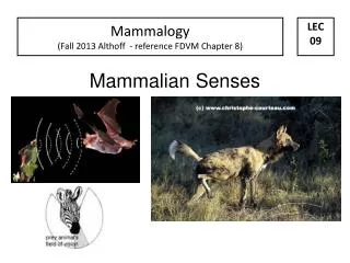
The Mammalian Brain. 9.3 The Central Nervous System p. 427 - 434. The Central Nervous System. Consists of the brain and the spinal cord. These organs are protected by: 1) skull and vertebrae 2) cerebrospinal fluid cushions the brain and spinal cord

E N D
The Mammalian Brain 9.3 The Central Nervous System p. 427 - 434
The Central Nervous System • Consists of the brain and the spinal cord. These organs are protected by: • 1) skull and vertebrae • 2) cerebrospinal fluid cushions the brain and spinal cord • 3) meninges are protective membranes that surround the CNS
The Spinal Cord • Contains interneurons that link the sensory and motor pathways. These neurons carry information to and from the brain. The spinal cord contains 31 segments, each of which has a pair of spinal nerves. • The spinal cord contains white matter (bundles of myelinated axons of sensory and motor neurons), and grey matter (unmyelinated interneurons and the dendrites of motor neurons).
Cerebrospinal fluid = surrounds CNS (brain and spinal cord); acts as a sock absorber and transport medium (nutrients, chemicals, removal of wastes, etc) is a connection between CNS and endocrine system
The Brain • The human brain contains three distinct areas: the forebrain, the midbrain, and the hindbrain. • is protected by the skull and meninges (a protective three layer thick membrane that surrounds the brain and spinal cord) • meninges controls which chemicals can ultimately reach the brain
The meninges (membrane) covers the surface of the cerebrum The most common symptoms of meningitis are headache and neck stiffness associated with fever Meningitis is diagnosed using a technique called lumbar puncture
The meninges is composed of 3 membranes; the dura mater, the tough outer membrane that adheres to the skull The arachnoid, the weblike middle layer that reabsorbs cerebrospinal fluid; And the pia mater, the innermost layer that contains many blood vessels and closely covers the brain and spinal cord
Which region is responsible for vision? Which is responsible for hearing?
Sections of the Brain Forebrain: • reason, intellect, memory, language, and personality • information on right side does not = info on left • generally on right (visual patterns or spatial awareness) • generally on left (verbal skills) • hemispheres are joined by a bundle of nerves called corpus collosum = allows communication between hemispheres
cerebrum is composed of 2 hemispheres What forms the largest part of the brain?
Why are we smarter than fish? We share a common ancestor However, our forebrains are much larger (cerebrum: speech, reasoning, memory, and personality)
“an elephant never forgets” The cerebrum (area of memory) is well developed in the elephant The elephants cerebrum is larger than humans
The increased surface area allows more nerve cells, which in turn allows for more learning and a greater range of behaviours. The grey matter of the human cortex is less than 5 mm thick, but in mass makes up more than 80% of the brain.
The central fissure extends from the top of each cerebral hemisphere to the lateral fissure
Midbrain: • located directly below cerebral cortex • relay center for eye and ear reflexes
http://outreach.mcb.harvard.edu/animations/brainanatomy.swf The Hindbrain:
Cerebellum What does convoluted mean? Damage to the cerebellum can lead to: loss of coordination of motor movement
brain stem basic attention, arousal, and consciousness
The frontal lobe: higher intellect, foresight and judgment, primary motor area and motor area for speech In 1890, psychiatrist Gottlieb Burckhardt removed pieces of the frontal lobes of six patients in a psychiatric hospital in Switzerland.
The parietal lobe: touch, temperature, and taste, and association areas for emotions, reading, and interpreting speech
Temporal: smell, hearing and auditory association areas
The corpus callosum contains nerve fibers that connect the right and left sides of the brain
The corpus callosum is white because it consists of myelinated nerve fibres. Sheep
The medulla oblongata controls heart and breathing rates and vasomotion (the dilation and constriction of blood vessels) to ensure blood is distributed more to active tissues than inactive ones. sheep
The pons is an important relay center for sensory and motor nerve fibers connecting the medulla oblongata and the cerebellum The pons also stimulates exhalation during prolonged inhalation of breath-holding.
The hypothalamus is an important center for the homeostatic regulation of several activities. It produces the hormones oxytocin and antidiuretic hormone secreted by the posterior pituitary gland. Other functions include the regulation of body temperature, water retention, appetitie, digestive secretions, sexual activity, and emotions such as fear and rage.
PRACTICE!!!1. List the four regions of the cerebral cortex and state the function of each. Frontal lobe: motor areas control movement of voluntary muscles (e.g., walking and speech). Association areas are linked to intellectual activities and personality. b) Temporal lobe: Sensory areas are associated with vision and hearing. Association areas are linked to memory and interpretation of sensory information.
c) Parietal lobe: Sensory areas are associated with touch and temperature awareness. Association areas have been linked to emotions and interpreting speech. d) Occipital lobe: Sensory areas are associated with vision. Association areas interpret visual information.
3. Name the different parts of the brain on page 434 and give functions. T- cerebrum: stores sensory information and initiates voluntary motor activities S-pons: acts as a relay station by sending nerve messages between the cerebellum and the medulla R- medulla oblongata: site of autonomic nerve control V- cerebellum: coordinates muscle movent
4. A physician makes an incision completely through the corpus callosum. How might this affect the patient? The right and left sides of the brain will not be able to communicate. “the left hand does not know what the right hand is doing” Has been considered as a treatment for severe cases of epilepsy
Phineas P. Gage (July 9?, 1823–May 21, 1860) was a railroad construction foreman now remembered for his incredible survival of an accident which drove a large iron rod through his head, destroying one or both of his frontal lobes.
Magnetic resonance imaging(MRI), or Nuclear magnetic resonance imaging (NMRI) Brain Anatomy and Function