Download
1 / 9
331 likes | 1.88k Views
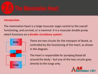
Body. Heart. Lungs. 2.6. The Mammalian Heart. Introduction The mammalian heart is a large muscular organ central to the overall functioning, and survival, or a mammal. It is a muscular double pump which functions on a double circulatory system .

E N D
Body Heart Lungs 2.6 The Mammalian Heart Introduction The mammalian heart is a large muscular organ central to the overall functioning, and survival, or a mammal. It is a muscular double pump which functions on a double circulatory system. There are two circuits for the transport of blood, as controlled by the functioning of the heart, as shown in the diagram. The heart is responsible for pumping blood all around the body – but one of the two circuits goes directly to the lungs only.
2.6 The Mammalian Heart • The Double Pump • The heart is responsible for two completely different processes which are linked together on the double circulatory system: • the right side pumps deoxygenatedblood to the lungs to be oxygenated • the left side pumps the oxygenated blood to the rest of the body • The heart squeezes the blood – which puts it under pressure. This in turn, forces the blood along arteries.
2.6 The Mammalian Heart The External Structure of the Heart The heart is made mainly of dark red muscle, which surrounds the two main pumping chambers: the ventricles. Above the ventricles are two thin-walled chambers: the atria(plural of atrium). There are coronary arteries which lay over the heart surface to provide the heart muscle itself with oxygenated blood. If blood flow to the heart is restricted it may cause anginaor a heart attack (myocardial infarction). There are a number of larger veins and arteries at the very top of the heart which carry the blood into and out of the heart.
2.6 The Mammalian Heart Internal Features of the Heart PRACTICE Name as many features of the heart, as shown in the diagram, as you can aorta vena cava pulmonary artery semilunar valve pulmonary vein right atrium left atrium right atrioventricular valve semilunar valve left atrioventricular valve right ventricle left ventricle papillary muscle ventricular septum tendinous cords
2.6 The Mammalian Heart Internal Features of the Heart The heart is split into four chambers: the two atria and two ventricles. Deoxygenated blood flows from the vena cava into the right atrium. Oxygenated blood flows from the lungs flows from the pulmonary vein into the left atrium. From the atria, blood flows down through atrioventricular valves (the left being called bicuspidand the right tricuspid) into the ventricles. These are pocket tissues which fill up with blood and remain closed whenever the ventricles contract. This ensures the blood flows upwards into the major arteries and not back into the atria.
2.6 The Mammalian Heart Internal Features of the Heart Inside the ventricles are tendinous cords, which attach the valves to the walls of the ventricle, preventing the valves from turning inside out, which would also allow the backflow of blood back into the atria. A wall of muscle (called the ventricular septum) separates the ventricles from each other. This ensures that the oxygenated blood in the left side of the heart and the deoxygenated blood in the right side are kept separate.
2.6 The Mammalian Heart Internal Features of the Heart Deoxygenated blood leaving the right ventricle flows into the pulmonary arteryleading to the lungs. Oxygenated blood leaving the left ventricle flows into the aorta. This carries blood to a wide series of arteries which supply the rest of the body with blood. At the base of the major arteries, where they exit the heart, are semilunar valves which prevent the blood returning the heart as the ventricles relax.
2.6 The Mammalian Heart Blood Pressure The muscle of each chamber contracts to create an increased pressure in the blood. The higher the pressure created in the heart, the further it will push the blood. The muscle of the atria wall is very thin. This is because their only function is to push the blood into the ventricles, so there is no need for a very high pressure.
2.6 The Mammalian Heart Blood Pressure The walls of the right ventricle are thicker. This enables it to pump the blood out of the heart. The left ventricle has walls which are even thicker than the right. Mainly, this is because the left ventricle needs to pump blood all around the body, whereas the right ventricle only needs to pump blood round to the lungs, which are situated alongside the heart in the chest cavity, so the distance it needs to be pumped is a lot smaller. Another reason is that the lungs contain a network of very fine capillaries which are in close contact with the alveoli. The alveoli walls are very thin, and therefore the capillaries are not supported and could therefore burst if there were a too high pressure.