Download

1 / 46
460 likes | 623 Views
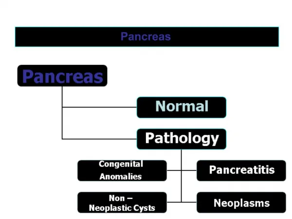
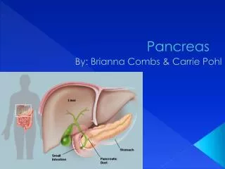
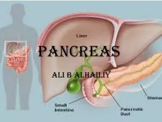
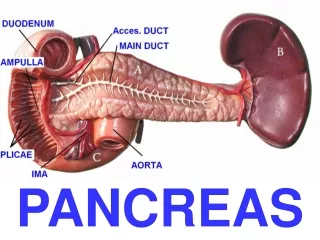
Pancreas. Oblong gland that lies posterior to the greater curvature of the stomach. Connected by ducts to the duodenum. Composed of clusters of glandular epithelial cells. Two main types of Pancreatic Cells: Pancreatic Islets-Islets of Langerhans (1%)

E N D
Pancreas • Oblong gland that lies posterior to the greater curvature of the stomach. • Connected by ducts to the duodenum. • Composed of clusters of glandular epithelial cells. • Two main types of Pancreatic Cells: • Pancreatic Islets-Islets of Langerhans (1%) • Hormones: insulin, glucagon, somatostatin • Acini Cells (99%) • Digestive pancreatic enzymes
Pancreatic Juice • Alkaline mixture of fluid and digestive enzymes from the acini cells. • Pancreatic Digestive Enzymes: • Pancreatic Amylase - CHO digestion • Pancreatic Lipase - Fat digestion • Chymotrypsin-Trypsin-Carboxypeptidase - Protein digestion • Nucleases - Nucleic Acid digestion • Regulated by the intestinal hormones: Secretin and Cholecystokinin.
Liver • Located just under the diaphragm on the right side of the body. • Largest organ of the abdominopelvic cavity. • Weighs about 1.4 kgs (3 lbs). • Nicknamed the chemical factory of the body. • Completely covered by the peritoneum and a dense layer of connective tissue beneath the peritoneum.
Anatomy of the Liver • Right Lobe - largest lobe of the liver. • Located on the lateral-right side of the body • Caudate Lobe - posterior portion of right lobe • Quadrate Lobe - inferior portion of right lobe • Left Lobe - smaller, medial lobe of the liver. • Falciform Ligament - separates the right and left lobes of the liver and anchors it to the diaphragm and anterior abdominal wall.
Lobules of the Liver • Smaller functional units of the liver. • Hepatocytes in the lobules produce and secrete a yellowish, brownish, or olive green liquid called BILE (1 quart daily). • Composed of bile salts and pigments, lecithin, and several ions. • pH of 7.6 - 8.6. • Excretory product and digestive secretion. • Assists in the breakdown of fat molecules (Emulsification). • Principle bile pigment is bilirubin.
Functions of the Liver • Metabolism of CHO, fats, and proteins • Removal of drugs and hormones • Excretion of bile • Synthesis of bile salts • Storage of vitamins, minerals, and food molecules • Phagocytosis of old, worn out RBCs and WBCs • Activation of Vitamin D
The Gallbladder • A pear shaped sac about 7 - 10 cm long. • Located on the bottom surface of the liver. • Stores and concentrates bile until needed by the small intestine for the emulsification of fat.
The Small Intestine • Duodenum - the beginning of the small intestine. Is attached to the stomach. • first 12 to 14 inches • Jejunum - the portion of the small intestine after the duodenum. • normally about 8 ft. long • Ileum - the final portion of the small intestine. • about 12 ft. long • ileocecal valve
Chemical Digestion of the Small Intestine • Very complex process with a series of chemical events that results in the breakdown of carbohydrates, fats, and proteins. • Result of the collective effort of pancreatic juice, bile, and intestinal juice. • Results in absorption - passage of digested nutrients into the blood or lymph.
Mechanisms to Increase Absorption by the Small Intestines • Folds in the intestinal walls of the mucosa layer of tissue . • (Plicae Circulares) • Villi arrangement of tissue of mucosa layer. • Lacteals - blood capillaries and lymphatic vessels associated with each villi • Microvilli arrangement of epithelial cells of the mucosa.
Absorption in the Small Intestine • 90% of absorption takes place within the small intestine. • other 10% occurs in the stomach and large intestine • Absorption of nutrients occurs through the villi by means of: • diffusion - facilitated diffusion • osmosis - active transport
Additional Components of the Small Intestine • Intestinal Juice - slightly alkaline secretion (pH 7.6) secreted by intestinal glands. • rapidly absorbed by the villi and provides a mechanism for absorption of substances in chyme • Peyer’s Patches - lymphatic glands of the small intestine. • Brunner’s Glands - mucus secreting glands of the small intestine.
Mechanical Digestion of the Small Intestine • Segmentation - localized contraction of muscles of the small intestine in areas containing food • rate of about 12 - 16 contractions/minute • sloshing of chyme back and forth within the intestinal lumen • Peristalsis - rhythmical contraction of muscles of the small intestines that propels chyme through the intestinal tract
The Large Intestine • About 1.5 m (5 ft) in length. • Cecum - beginning of the large intestine • Vermiform Appendix • Colon - large tube-like portion of large intestine • Ascending Colon - Transverse Colon • Descending Colon - Sigmoid Colon • Rectum • Anal Canal --->Anus
Functions of the Large Intestine • Completes absorption • Teabsorption of water • Manufacture certain vitamins • Formation of feces • Expulsion of feces from the body
Histology of the Large Intestine • Walls of the large intestine contain no villi or permanent circular folds in the mucosa layer. • Epithelial tissue layer contain numerous goblet cells (secretes mucus). • Lubricates the colonic contents as it passes through the large intestine.
Haustra - series of characteristic pouch like structures that run the entire length of the colon. • Taenia Coli - bands of smooth muscle that are arranged longitudinally along the length of the colon. • Epiplotic Appendages - small fat filled appendages suspended from the Taenia Coli. • Anal Columns - parallel ridges of mucosa in the anal canal which reduces friction with feces during defecation.
Mechanical Digestion in the Large Intestine • Haustral Churning - the relaxation and contraction of the individual segments of the colon. • Peristalsis - rhythmical contraction of the colon that moves the contents along through the length of the colon. • Mass Peristalsis - a strong peristaltic wave that begins about the middle of the transverse colon and drives the colonic contents into the rectum.
Chemical Digestion in the Large Intestine • last stage of digestion • due to bacterial action in the large intestine • bacteria ferment any remaining carbohydrates and release hydrogen, carbon dioxide, and methane gas • also converts any remaining proteins into amino acids • absorbs any remaining water and electrolytes
Feces Formation in the Large Intestine • by the time chyme has remained in the large intestine for 3 - 10 hours it has become a solid or semi-solid and is known as FECES • consists of water, inorganic salts, sloughed off epithelial cells, products from bacterial decomposition, and indigestible parts of food Defecation • The emptying of the rectum. • Diarrhea - frequent defecation of liquid feces. • Constipation - infrequent or difficult defecation. Stools are hard and dry.
Digestive System Diseases and Homeostatic Imbalances Cirrhosis of the Liver Distorted or scarred liver tissue due to chronic inflammation. Commonly caused by hepatitis, chemical exposure, parasites, and alcoholism. Symptoms include: jaundice, bleeding, edema, and increased sensitivity to drugs and chemicals.
Appendicitis • Inflammation of the vermiform appendix. • May be caused by an obstruction of the lumen of the appendix by fecal material, a foreign body, stenosis, a kinking of the organ, or carcinoma. Gall Stones • Crystallization of bile in the gallbladder. • Can block the bile duct causing intense pain. • Usually treated with gall stone dissolving drugs, lithotripsy, or surgery.
Tumors of the Digestive Tract • Can occur in all areas of the G.I. tract. • Can be malignant or benign. Often no smptoms. • Colorectal Cancer • 3rd most common cause of cancer for both males and females. • Overall mortality rate is over 60%. • Factors contributing to colorectal cancer include genetic predisposition, diet high in fat, protein, insufficient dietary fiber, and low calcium and selenium in the diet.
Hepatitis • Inflammation of the liver. • Can be caused by viruses, drugs, and certain chemicals including steroids and alcohol. • Many different types of Hepatitis including: • Hepatitis A (Infectious Hepatitis) • Hepatitis B (Serum Hepatitis) • Non A Non B (NANB) Hepatitis
Hepatitis A • Infectious hepatitis • Caused by Hepatitis A virus • Spread by fecal contamination of food, clothing, toys, eating utensils, etc. • Generally a mild disease of children and young adults • Characterized by anorexia, malaise, jaundice, nausea, diarrhea, fever, and chills
Hepatitis B • Serum hepatitis • Caused by the Hepatitis B virus • Transmitted by sexual contact, contaminated syringes, transfusion equipment, saliva, tears, and puncture wounds in the skin • Can produce cirrhosis and possibly cancer of the liver Non A Non B Hepatitis • Other types of hepatitis that can not be traced to either the A or B virus (up to about virus H in the alphabet). • Often spread by blood transfusions. • Can cause cirrhosis and possibly cancer of the liver.
Obesity • Clinically classified as obese if: • > 30% of projected body weight as determined height and frame size • doesn’t factor in Body Composition • Currently over 50% of U.S. population is clinically classified as obese. • 14% of all male cancers linked to obesity 20% of all female cancers linked to obesity Diabetes mellitus rates continue to rise dramatically in the U.S.
U.S. surgeon general has said obesity is the second most serious threat to the health of Americans. • A serious risk factor for: • Heart Disease - Diabetes • Hypertension - Cancers • Respiratory Disorders - Joint Problems • Endocrine Disorders • Gastrointestinal Disorders • Urinary and Reproductive System Disorders
Peptic Ulcers • Crater-like lesions that develop in the gastrointestinal tract. • Gastric Ulcers ---> Stomach • Duodenal Ulcers ---> Duodenum • Commonly caused by hypersecretion of gastric juices and acids. • Contributing factors include: stress, cigarette smoking, certain foods, some medications, and bacterial infections.