PANCREAS
PANCREAS. Dr Jamila Elmedany & Dr Saeed Vohra. OBJECTIVES. By the end of this lecture the student should be able to : Describe the anatomical view of the pancreas regarding ; location, parts relations, ducts Arterial supply & Venous drainage Describe the nerve supply and lymph drainage.

PANCREAS
E N D
Presentation Transcript
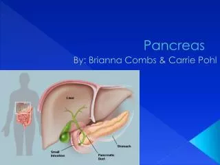
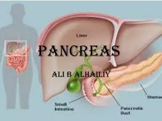
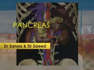
PANCREAS Dr JamilaElmedany & Dr SaeedVohra
OBJECTIVES • By the end of this lecture the student should be able to: • Describe the anatomical view of the pancreas regarding ; location, parts relations, ducts • Arterial supply & Venous drainage • Describe the nerve supply and lymph drainage
PANCREAS It is an elongated soft pinkish structure (60-100) gram in weight & (6-10) inch in length It isLobulated? Becauseit is surrounded by a fibrous tissue capsule from which septa pass into the gland and divide it into lobes. The lobes are divided into lobules.
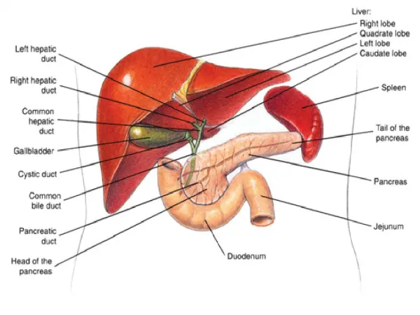
LOCATION • It is a Retro-Peritoneal structure. • It lies on the posterior abdominal wall in the: Epigastrium & Left upper quadrant of the abdomen. • It extends in a transverse oblique direction at the transpyloric plane (1st lumbar vertebral) from the concavity of the duodenum on the right to the spleen on the left.
PARTS • It is divided into: • Head, Neck, Body and Tail. • Because of its oblique direction the tail is higher than the head.
Head of Pancreas • It is disc shaped • Lies within the concavity of the duodenum • Related to the 2nd and 3rd portions of the duodenum. • On the right, it emerges into the neck. • On the left, it Includes Uncinate Process ( an extension of the lower part of the head behind the superior mesenteric vessels)
Structures Posterior to the Head: (1) Bile Duct runs downwards and may be embedded in it.? (2) IVCruns upwards.
Neck of Pancreas • It is the constricted portion connecting the head & body of pancreas • It lies in front of: • Aorta • Origin of Superior Mesenteric artery • the confluence of the Portal Vein • Its antero-superior surface supports the pylorus of the stomach • The superior mesenteric vessels emerge from its inferior border
Body of Pancreas • It runs upward and to the left. • It is triangular in cross section. • TheSplenicVein is embedded in its post. Surface • TheSplenicArtery runs to the left along the upper border of the pancreas.
Tail of Pancreas A narrow, short segment lies at the level of the 12th thoracic vertebra Ends within the splenichilum Lies in the Splenicorenalligament Anteriorly, related to: splenic flexure of colon May be injured during Splenectomy
RELATIONS OF PANCREAS • Anterior to (body & tail): • Stomachseparated from by lesser sac • Transverse colon & transverse mesocolon
Posterior to (body & tail) : • LeftPsoas muscle • Left Adrenal gland • Left Renal vessels • Upper 1/3rd of Left kidney • Hilum of the spleen.
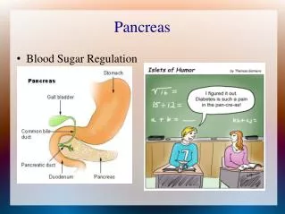
FUNCTIONS Exocrine and Endocrine The Exocrine portion: Small ducts arise from the lobules and enter the main pancreatic duct (it begins in the tail), and passes through the body and head where it meets the bile duct. The Endocrine portion: (Islets of Langerhans) produce insulin & glucagon.
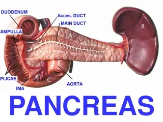
Pancreatic DUCTS • Main P duct : • Joins common bile duct &they open into a small hepatopancreaticampulla in the duodenal wall (Ampulla of Vater). • The ampulla opens into the lumen of the duodenum through (Major Duodenal Papilla).
Accessory P duct (of Santorini) Drains superior portion of the head • It empties separately into 2nd portion of duodenum at (minor duodenal papilla)
ARTERIAL SUPPLY • Celiac trunk, Superior mesenteric & Splenic arteries Celiac T CHA • Gastroduodenal • Superior pancreaticoduodenal SMA Inferior pancreaticoduodenal TO HEAD Splenic Asupplies the Body and Tail of pancreas by about 10 branches R gastric Hepatic
VENOUS DRAINAGE • Anterior and posterior arcades drain head and the body • Splenic vein drains the body and tail • Ultimately, end into Portal Vein
LYMPHATIC DRAINAGE • Rich network drains into nodes along the upper border of the pancreas • Ultimately the efferent vessels drain into the Celiac nodes. • Lymph vessels from the region of the Head pass to • Superior Mesenteric nodes • (Important ?)
NERVE SUPPLY • Sympatheticfrom the splanchnicnerves , they have a predominantly inhibitory effect • Parasympatheticfrom the Vagus, • they stimulate both exocrine and endocrine secretions