Download

1 / 35
360 likes | 720 Views
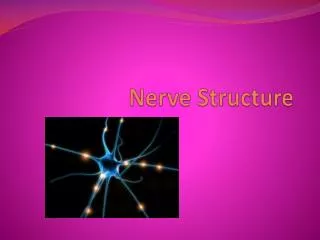
NERVE TISSUE. STRUCTURALLY NERVOUS TISSUE IS OF TWO TYPES. nerve cells, or neurons, which usually show numerous long processes are responsible for the reception, transmission, processing of stimuli; the triggering of certain cell activities;

E N D
STRUCTURALLY NERVOUS TISSUE IS OF TWO TYPES nerve cells, or neurons, which usually show numerous long processes • are responsible for the reception, transmission, processing of stimuli; • the triggering of certain cell activities; • the release of neurotransmitters and other informational molecules.
Glial cells (Gr. glia, glue), which have short processes, • support and protect neurons, • participate in neural activity, • neural nutrition, • defense processes of the central nervous system
Parts of neuron Most neurons consist of three parts • the dendrites, which are multiple elongated processes specialized in receiving stimuli from the environment, sensory epithelial cells, or other neurons;
the cell body, or perikaryon (Gr. peri, around, + karyon, nucleus), which is the trophic center for the whole nerve cell and is also receptive to stimuli;
The cell body contains a highly developed rough endoplasmic reticulum. When appropriate stains are used, rough endoplasmic reticulum and free ribosomes appear under the light microscope as basophilic granular areas called Nissl bodies
axon , which is a single process specialized in generating or conducting nerve impulses to other cells (nerve, muscle, and gland cells).
All axons originate from a short pyramid-shaped region, the axon hillock, that usually arises from the perikaryon .
The plasma membrane of the axon is called the axolemma (axon + Gr. eilema, sheath); its contents are known as axoplasm.
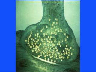
The distal portion of the axon is usually branched and constitutes the terminal arborization. • .
Each branch of this arborization terminates on the next cell in dilatations called end bulbs (boutons), which interact with other neurons or nonnerve cells, forming structures called synapses.
Glial Cells & Neuronal Activity • Glial cells are 10 times more abundant in the mammalian brain than neurons; they surround both cell bodies and their axonal and dendritic processes that occupy the interneuronal spaces.
Oligodendrocytes • Oligodendrocytes (Gr. oligos, small, + dendron + kytos, cell) produce the myelin sheath that provides the electrical insulation of neurons in the central nervous system
Schwann Cells • Schwann cells have the same function as oligodendrocytes but are located around axons in the peripheral nervous system.
Astrocytes • Astrocytes (Gr. astron, star, + kytos) are star-shaped cells with multiple radiating processes. Astrocytes bind neurons to capillaries and to the pia mater (a thin connective tissue that covers the central nervous system). • Astrocytes with few long processes are called fibrous astrocytes and are located in the white matter; • protoplasmic astrocytes, with many short-branched processes, are found in the gray matt
Ependymal Cells • Ependymal cells are low columnar epithelial cells lining the ventricles of the brain and central canal of the spinal cord.
Microglia • Microglia (Gr. micros, small, + glia) are small elongated cells with short irregular processes. Microglia, are derived from precursor cells in the bone marrow. • They are involved with inflammation and repair in the adult central nervous system
Organization of nervous tissue in Central Nervous System • The central nervous system consists of the cerebrum, cerebellum, and spinal cord. It has almost no connective tissue and is therefore a relatively soft, gel-like organ. • When sectioned, the cerebrum, cerebellum, and spinal cord show regions that are white (white matter) and that are gray (gray matter).
The main component of white matter is myelinated axons and the myelin-producing oligodendrocytes. White matter does not contain neuronal cell bodies.
Gray matter contains neuronal cell bodies, dendrites, and the initial unmyelinated portions of axons and glial cells.
Peripheral Nervous System • main components of the peripheral nervous system are the nerves, ganglia, and nerve endings.
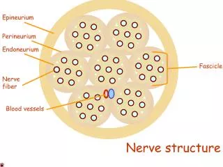
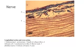
Nerves are bundles of nerve fibers surrounded by connective tissue sheaths. • Nerve fibers consist of axons . • Groups of nerve fibers constitute the tracts of the brain, spinal cord, and peripheral nerves.
Nerves have an external fibrous coat of dense connective tissue called epineurium, which also fills the space between the bundles of nerve fibers. Each bundle is surrounded by the perineurium, a sleeve formed by layers of flattened epitheliumlike cells.
Ganglia • Ganglia are discrete aggregations of neuronal cell bodies located outside the CNS • The direction of the nerve impulse determines whether the ganglion will be a sensory or an autonomic ganglion.
Sensory Ganglia • Sensory ganglia receive afferent impulses that go to the central nervous system. • Two types of sensory ganglia exist. Some are associated with cranial nerves (cranial ganglia); • associated with the dorsal root of the spinal nerves and are called spinal ganglia.
Sensory Ganglia • The whole ganglion is encapsulated by condensed supporting tissue which is continuous with the perineurial and epineurial sheaths of the associated peripheral nerve.
Autonomic Ganglia • These ganglia are devoid of connective tissue capsules, and their cells are supported by the stroma of the organ in which they are found.
The ganglion cells are multipolar and thus more widely spaced, . • the nuclei of the ganglion cells tend to be eccentrically located .