Download
1 / 34
340 likes | 359 Views
Learn about the composition and functions of epithelial, muscle, nerve, and connective tissues in the human body and their microscopic structures.

E N D
EPITHELIAL TISSUE MUSCLE TISSUE NERVE TISSUE CONNECTIVE TISSUE
EPITHELIAL TISSUE • Epithelial tissue or epithelium is composed of one or more layers of cells, arranged next to each other without any space between them. • This tissue covers the external surface of body, forming the skin, and lines cavities and tracts such as the stomach and the intestines.
Cell of epithelial tissue Cell nucleus •The epithelial cells are arranged in many layers, which is why it is called pluristratified epithelial tissue. • The nucleus of the cell can be seen well, as it has been stained a dark purple colour. EPITHELIAL TISSUE •Image of epithelial tissue taken from an optical microscope.
Cell of epithelial tissue Cell nucleus • The cells are prism-like in shape and their nuclei have been stained a dark blue colour. EPITHELIAL TISSUE • Image of epithelial tissue taken from an optical microscope. • The epithelial cells are arranged in two layers, but they are not aligned, so it is called pseudostratified epithelial tissue
EPITHELIAL TISSUE MUSCLE TISSUE NERVE TISSUE CONNECTIVE TISSUE
MUSCLE TISSUE • Muscle tissue is composed of elongated cells, the muscle fibres. These fibres are contractile, which means they contract in response to stimuli. • The muscle tissue forms the muscles of the locomotor system (skeletal muscles), the heart (cardiac muscle or myocardium) and the walls of some organs (smooth muscle).
Nuclei of the muscle fibres • The muscle fibres are elongated, cylindrical and striated (arranged in parallel). •The nuclei of the muscle fibres are elongated and found along the sides of the fibres. Muscle fibres MUSCLE TISSUE •Image of muscle tissue taken using an optical microscope. •We can see a longitudinal section of skeletal muscle tissue.
Nuclei of muscle fibres •The muscle fibres, cut transversally, have a blocky structure. • The nuclei can be seen around the sides of each fibre. Muscle fibre MUSCLE TISSUE • Image of muscle tissue taken using an optical microscope. •A cross section of skeletal muscle tissue can be seen.
EPITHELIAL TISSUE MUSCLE TISSUE NERVE TISSUE CONNECTIVE TISSUE
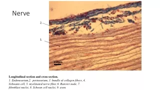
NERVE TISSUE • Nerve tissue is made up of neurons, cells capable of capturing and responding to stimuli, and of controlling the activity of the organism.
Neuron •In the image, we can see two neurons. Nucleus of the neuron •The largest neuron has a polygonal (or star) form and at the nucleus is in the centre. Prolongations •The body of the neuron has five prolongations. NERVE TISSUE •Image of nerve tissue taking using an optical microscope.
Neuron Nucleus of the neuron • The bodies of the neurons have a polygonal form with the nuclei at the centre. Prolongations •The prolongations of these neurons are very long. NERVE TISSUE •Image of nerve tissue taken under an optical microscope. •In the image, we can see two neurons.
EPITHELIAL TISSUE MUSCLE TISSUE NERVE TISSUE CONNECTIVE TISSUE
CONNECTIVE TISSUE • Connective tissue is composed of cells separated by an intercellular substance called the matrix. Their function in the body is to connect and support. • There are various types of connective tissue: conjunctive tissue, cartilaginous tissue, adipose tissue, bone tissue and blood tissue.
CONNECTIVE TISSUE CONJUNCTIVE CARTILAGINOUS ADIPOSE BONE BLOOD
CONJUNCTIVE TISSUE • Conjunctive tissue is a type of connective tissue which joins the other tissues together.
Liver cells • The dark round points are the liver cells. •The lines are fibres, which form a network that provides support to the organ, in this case the liver. Fibres CONJUNCTIVE TISSUE • Image of conjunctive tissue taken using an optical microscope
CONNECTIVE TISSUE CONJUNCTIVE CARTILAGINOUS ADIPOSE BONE BLOOD
CARTILAGINOUS TISSUE • This is a type of connective tissue which forms part of the skeleton and provides support to the soft parts of the body.
Matrix of the cartilaginous tissue • The light blue colour (background) is to the intercellular matrix which surrounds the cells of the cartilaginous tissue, which are called chondrocytes. • The chondrocytes have been stained a dark blue colour. Chondrocytes CARTILAGINOUS TISSUE •Image of cartilaginous tissue from the trachea,taken using an optical microscope.
CONNECTIVE TISSUE CONJUNCTIVE CARTILAGINOUS ADIPOSE BONE BLOOD
ADIPOSE TISSUE • Adipose tissue is a type of connective tissue that makes up the organism’s greatest energy reserve and provides thermal insulation. It is found under the skin.
Adipose tissue cell Lipid droplet •The cells that form this tissue are round in shape. •The cells have a single lipid droplet, which is so large that it occupies almost the entire cell. ADIPOSE TISSUE •Image of adipose tissue taking using an optical microscope. •This is an example of white adipose tissue.
Adipose tissue cell Lipid droplets •The cells that make up this tissue are polygonal in shape. Cell nucleus •Unlike white adipose tissue, the cells of this tissue contain many lipid droplets. •The nucleus is found on one side of the cell. ADIPOSE TISSUE •Image of adipose tissue taken using an optical microscope. •This is an example of brown adipose tissue, which is scarce in human beings and typical in animals that hibernate.
CONNECTIVE TISSUE CONJUNCTIVE CARTILAGINOUS ADIPOSE BONE BLOOD
BONE TISSUE • Bone tissue is a type of connective tissue that provides support to the organism and protects the vital organs.
Osteon • In the centre of the image, two structures called osteons can be seen. •The osteons are concentric layers of bone tissues surrounding a central channel. Central channel of the osteon •In the osteons, we can see black spots, which are the spaces containing the bone cells. Black spots BONE TISSUE • Image of bone tissue taken using an optical microscope.
CONNECTIVE TISSUE CONJUNCTIVE CARTILAGINOUS ADIPOSE BONE BLOOD
BLOOD TISSUE • In this type of connective tissue the liquid matrix is called plasma. • Suspended in the plasma are the blood cells (the red blood cells,white blood cells and platelets).
Blood plasma •The liquid matrix, in this case the plasma, surroundins the cells. White blood cells •The majority of blood tissue is made up of red blood cells. •In the image, we can also see white blood cells, which have been stained purple. Red blood cells BLOOD TISSUE •Photograph of a sample of human blood taken using an optical microscope.
Red blood cell White blood cell -The red blood cells are disc-like in shape and do not have nuclei. Nucleus -The nuclei of the white blood cells are brightly coloured purple. This nucleus has a very strange shape with small lobes attached to each other. BLOOD TISSUE •Previous image seen in greater magnification. •When the image is enlarged, we can the cells of this tissue in more detail:
CONNECTIVE TISSUE CONJUNCTIVE CARTILAGINOUS ADIPOSE BONE BLOOD
EPITHELIAL TISSUE MUSCLE TISSUE NERVE TISSUE CONNECTIVE TISSUE