Download
1 / 16
160 likes | 265 Views
Light-activated ion channels for remote control of neuronal firing. Banghart, M. et al Choi, Yu Yong. Background. Techniques for controlling neural activity have considerable limitations

E N D
Light-activated ion channels for remote control of neuronal firing Banghart, M. et al Choi, Yu Yong
Background • Techniques for controlling neural activity have considerable limitations • Traditional electrical and neural chemical methods requires invasive electrodes and chemical delivery systems that cannot control patterns of activity in densely packed neural tissue • Optical techniques utilizing caged neurotransmitters are less invasive and more precise, but reversal of the effects of the uncaged transmitter is limited by its diffusion kinetics • Exogenous expression of genes encoding ion channels has been used to influence electrical activity in specific neurons, but the onset and reversal of gene expression is slow • Photic regulation has been conferred on neurons by introducing a rhodopsin-based signal transduction-based cascade, This technique requires coordinated exogenous expression of three different genes and produces light responses that can be slow in onset and offset and variable in different neurons, possibly because the nature of the native ion channels that are regulated by the cascade can vary between neurons • This light-activated ion channels allow rapid, remote, and noninvasive control. Because the light-activated gate is covalently linked to the ion channel, and because the ion-channel is integral to the neuronal cell membrane, control over individual neurons is spatially accurate and does not rely on a diffusible ligand. In addition, the gate can be reversly photo-switched, allowing recurrent control of neural activity
Rod cell • Figure 11.7. Details of phototransduction in rod photoreceptors. (A) The molecular structure of rhodopsin, the pigment in rods. (B) The second messenger cascade of phototransduction. Light stimulation of rhodopsin in the receptor disks leads to the activation of a G-protein (transducin), which in turn activates a phosphodiesterase (PDE). The phosphodiesterase hydrolyzes cGMP, reducing its concentration in the outer segment and leading to the closure of sodium channels in the outer segment membrane
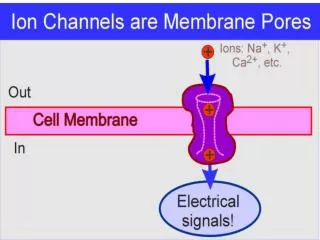
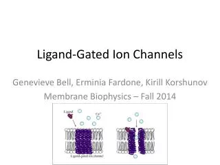
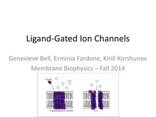
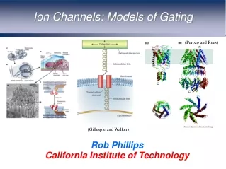
ion channels • Three physical models for the opening and closing of ion channels • A localized conformational change occurs in one region of the channel • A generalized structural change occurs along the length of the channel • A blocking particle swings into and out of the channel mouth • Types of • Ligand-gated • Voltage-gated • Stretch or pressure-gated
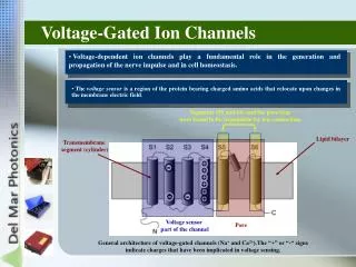
Shaker K+ channel • Typical voltage-gated K+ channels is an assembly of four identical (or similar) transmembrane subunits surrounding a central pore. Each subunit has six transmembrane (S1-S6), with both N- and C- termini on the intracellular side of the membrane (top left panel). The narrowest part of the pore, the selectivity filter, is formed by a loop between S5 and S6; the voltage sensor includes the S4 region with its multiple positive charges (Yellen, G., 2002. Nature )
Shaker K+ channel • Liu, Y. et al. 1997. Neuron
Shaker K+ channel • Blaustein, R. et al. 2000. Nat. Struct. Biol.
Patch Clamp • http://www.iac-usnc.org/Methods/wholecell/equipment.html Planar patch clamp
Patch Clamp • Note that only the on-cell patch and the whole-cell patch are associated with the complete neuron. The others are only concerned with a portion of membrane. Only the whole-cell approach actually accesses one of the plasmas within the neuron directly. In the case of the on-cell approach, the impedance of the membrane must be considered in the overall test set design. Planar patch clamp
Photocontrol of MAL-AZO-QA-modified Shaker channels in X. laevis oocytes
Discussion • By combining synthetic chemistry and protein mutagenesis, the Shaker channel has been reengineered • This technique lies in its spatial and temporal accuracy, its noninvasiveness and its reversibility. • and may provide an effective means for controlling the activity of specific neurons downstream from sites of neural damage or degeneration • and may be used to restore light-regulated activity in healthy retinal neurons after degeneration of rods and cones, the native phtoreceptors • and may have application in fields of nanotechnology, bioelectronics and material sciences