Download
1 / 52
550 likes | 830 Views
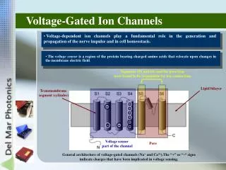
Second Messenger Activated Ion Channels: TRP Channels. Lindsey Biggs, Greg Loney Oct, 2011. Transient Receptor Potential (TRP) family of cation channels Drosophila trp mutation – resulted in transient response to light Typically flux Na + and/or Ca ++

E N D
Second Messenger Activated Ion Channels: TRP Channels Lindsey Biggs, Greg Loney Oct, 2011
Transient Receptor Potential (TRP) family of cation channels • Drosophila trpmutation – resulted in transient response to light • Typically flux Na+ and/or Ca++ • Single AA substitution alters cation selectivity • Often referred to as store operated channels or ‘SOCs’ • Expressed in both excitable and nonexcitable cells • Ubiquitouschannel family with various methods of activation: • Ligand (chemosensation) • Thermal (temperature) • Mechanoreceptor (somatosensation) O’Neil, 2011
Group 1 Group 2 TRPC TRPV TRPM TRPA TRPN Montell, 2011 Legend: Green = Ankyrin repeats Pink = Coiled coil domain Blue = TRP domain “P” = Pore domain Total number of channels expressed by exemplary species: Worms – 17 Flies – 13 Mice – 28 Humans – 27
Group 1 Group 2 TRPC TRPV TRPM TRPA TRPN Ankyrin –Present on NTD of TRP channels, typically 33 AA residues per repeat. Generally labeled as protein-protein interaction motifs and anchoring. Bind ATP, PLC, etc and may be important for channel sensing and gating properties TRP box – Located on CTD of S6, potentially important for channel closing/inactivation (?). Includes the invariant EWKFAR sequence that is conserved in all Trp proteins (?).
TRP channels are activated by signal transduction pathways. For a cell at rest, extracellular to intracellular ratios of Ca++ are ~20,000:1. It is likely that Ca++ modulates all TRP channels in some way, either directly or indirectly. TRP channels (e.g. TRPV1) are inhibited by PIP2 until it is hydrolyzed by PLC. PIP2 interacts with positively charged areas of channel (TRP box). Some channels (TRPM5) may be SOCs. Clapham, 2003
Three theories of SOCs: • Direct link between ER and channel • Putative second messenger • Fusion of vesicles containing SOC TRP channels Clapham, 2003
In vivo identification and manipulation of the Ca2+ selectivity filter in the drosophila transient receptor potential family Liu, Wang, Postma, Obukhov, Montell, Hardie 2007
Methods • Trpnull mutants demonstrate a transient LIC and sever retinal degeneration. • Generated mutant flies so that TRP was the only light sensitive channel present. TRPL mutants demonstrate highly selective Ca2+ current. • Asp621 and Asp626 are the only acidic residues unique to TRP sequence and are substituted in trpl mutants • Mutant flies were back-crossed to generate three different point mutated flies at Asp621 and one at Asp626 • Whole cell recordings were conducted from dissocatedommatidia
Figure 1: • Sequence homology between WT (trp) and trpl mutants • Protein expression amongst WT and mutants c. Immunolocalization of TRP channel – targeted to rhabdomeres
Figure 2: Reversal potentials obtained in control and biionic conditions (cation:Cs+) for flies expressing WT channels and point-mutated channels. Brief flashes were presented to ommatidia held at 10mv holding potential steps
Figure 3: Similar reversal potentials obtained under biionic conditions (cation:Cs+) for flies expressing WT channels and point-mutated channels in which the charge of the putative pore residue was titrated. Heteromultimers demonstrated intermediate Erev. Residues are plotted in order of decreasing charge
Figure 4: Reversal potentials obtained under biionic conditions for various mutants as a condition of cation size (A) and the inferred permeability ratios for these same cations
Figure 5: • Top trace is control noise present before channel activation. Bottom trace is steady-state noise after activation • Using both the control noise (top trace) and measured reversal potentials, data were fit to an equation in order to estimate the amount of single channel conductance • Quantum bumps (single absorbed photons)
Figure 6: • Inhibition of LIC as a function of Mg2+ in a Ca2+ free bath • I-V relationship of LIC. Asp626 mutants demonstrated a rectification current similar to WTs. The slope of the conductance between -10 and 40mv was lower in WT, presumably due to voltage dependent Mg2+ block (Ringer’s solution) • Demonstration of channel blockade by La3+ (left) and ruthenium red (right) - both polyvalent ions. La3+ presumably utilizes a distinct residue
Figure 7: A-D. Traces from WT and various mutants demonstrating the waveforms of flash induced LIC in both baths that contain Ca2+ and those that do not. E. Time-to-peak and time-to-decay. Note AspD621G mutants’ kinetics were not at all Ca2+ dependent. Often referred to as the fastest known G-protein coupled signaling cascade.
Figure 8: A-D. Responses to maintained illumination in WT and mutants. Asp621 mutants’ responses quickly decayed to baseline thus mimicking the original trp null mutants. E. Kir2.1 responses in WT and mutants. Kir2.1 is inwardly-rectifying K+ channel that is PIP2 dependent.
Summary • First demonstration, in vivo, that TRP is a pore forming channel that is selectively permeable to Ca2+ . • Altering a single amino acid residue in the putative selectively filter abolished Ca2+ selectivity and resulted in a phenotype that was indistinguishable from the original trp null mutant. • All that work summed in two points….thats why its in J Neuro…
Requirement of calcium-activated chloride channels in the activation of mouse vomeronasal neurons SangSeong Kim, Limei Ma, C. Ron Yu, 2011.
Review of TRPC2 in the accessory olfactory system • TRPC2 is found in all vomeronasal sensory neurons, but not in the main olfactory bulb or brain. • Located at the dendritic tip of the VSNs. • Field potential and extracellular recordings showed that responses in TRPC2 -/- mice to urine components were absent or diminished. • TRPC2 -/-mice show behavioral abnormalities: • Deficit s in male-male aggression and express subordinate behavior. (Also, seen in females). • Abnormalities in sexual behavior; increased male-male mounting, but normal mating with females. F. Zufall, 2005. Review. See for references.
TRPC2 is gated by DAG http://www.springerimages.com/Images/LifeSciences/1-10.1007_s00424-005-1432-4-9
Conditions used to study Cl- current Control: Control/mimicking natural conditions: Intracellular solution: contained Cl- Extracellular solution: also contained Cl- Expected effect upon channel opening: Cl- flow out of cell, resulting in an inward current flow (expulsion of negative current). Figure 1
Conditions used to study Cl- current Control: Control: MSF-: Methanesulfonate (MSF-): Non-permeable anion subsituted for Cl- in intracellular solution. Intracellular: very low Cl- levels Extracellular: High/normal Cl- Expected effect upon channel opening: very little to no Cl= mediated inward current. Figure 1
Conditions used to study Cl- current Low e.c. chloride: Control: MSF-: Figure 1 Gluconate substitution: Substitue non-permeable gluconate in place of chloride in extracellular solution. Intracellular: High/control Cl- levels Extracellular: Low Cl- levels Expected effect upon channel opening: large outflow of Cl- anions, resulting in a large inward current.
Urine activated chloride conductance in VNO neurons • As expected, altering intracellular (I.C.) chloride reduced the inward current. • Niflumic acid: • Cl- channel blocker. • Treatment reduced the induced inward current by 70%, similar to MSF- condition. Figure 1
VNO neuron sensitivity to small currents • Amplitude of inward current was very low, ranging from 2-12 pA. This is 10-100 times lower than for olfactory neurons, but this is supported by data from field recordings, showing 10-100 fold differences between MOE and VNO epithilium. Figure 1
Urine-activated Cl- current requires extracellular Ca2+ • E.C. calcium levels were then altered to determine the role of Ca2+ in these Cl- currents. • With less Ca2+ in the extracellular fluid, there were decreases in the urine-evoked inward currents, thus Ca2+ is necessary for urine-evoked currents. Figure 2
Urine-activated Cl- current requires intracellular Ca2+ • EGTA and BAPTA (Ca2+ chelating agents) were used in I.C. solutions to decrease I.C. Ca2+. • With low I.C. Ca2+, there was a reduction in the effect of E.C. Cl- ion substitution. Figure 2
Activation of Cl- current in TRPC2 -/- Mice • With low I.C. Cl-, there is no inward current, thus, all current seen in TRPC2 -/- mice is via Cl- channels. • In TRPC2 -/- mice, 80% of urine-evoked current was conserved, therefore, a TRPC2 independent mechanism is responsible for activation of CACC. Black: Cl- current Gray: non-Cl- current Figure 3
Intracellular Ca2+ release activates CACC • Since TRPC2 was the only TRP channel found in VNO neurons, they hypothesized that CACC was being activated by release of Ca2+ from intracellular stores. • In the presence of Ruthenium Red (RR, blocks IP3 mediated Ca2+ release) and Thapsigargin (TG, depletes I.C. Ca2+ stores), urine-induced currents were reduced. • In the MSF- condition where Cl- currents are not present, the effect of RR and TG are eliminated. • In TRPC2 -/- mice, RR and TG completely block urine-evoked response of VNO neuron.
TRPC2 KO doesn’t alter resting potential. • Extracellular recordings so as not to disturb I.C. Ca2+. • Urine application increased spiking rate in WT and KO (less firing in KO overall). • Block Cl-(NA or SITS)=blocked urine-induced spike increase. • TRPC2 channels alone are not sufficient to excite VNO neurons. WT= square KO= circle Conclusion: CACC are required for responses to urine.
Discussion/Conclusions • Cl- current contributes significantly to urine-evoked VNO responses. • This current is sufficient to drive changes in spiking rate in WT and TRPC2 -/-. • CACC currents were able to mediate pheromone activation independently of TRPC2 (Need to think about this conclusion). • Unlike the MOS, in which KO of CNG channel eliminates Cl- current, thus the animal is anosmic, only a portion of the inward current is carried by TRPC2, and TRPC2 influx of Ca2+. • Ca2+ influx via activation of TRPC2, and Ca2+ release from I.C. stores act synergistically to activate the Cl- conductance, but VNO neurons can be excited without TRPC2. • TRPC2 -/- behavioral phenotype may be explained by reduced VNO function and selective loss of the basal layer of VNO neurons (Go pathway).
Essential Role for TRPC5 in Amygdala Function and Fear-Related Behavior Antonio Riccio, et. al., 2009
Background: Fear circuitry • Lateral amygdala (LA) is the input center for auditory, somatosensory, visual, olfactory and taste systems. • Central amygdala(CeA) is the output center for fear conditioning circuitry.
Background: Methods- measures of anxiety • Elevated Plus Maze: Time spent and number of entries into open and closed entries. • More time/entries into open arm= less anxiety • Open Field test: Measure number of entries and time spent in center of the open field. • More time/more entries into center= less anxiety. • Social interaction: place animal in center cage and record time spent in area with novel and familiar conspecifics. Also, record nose contacts. http://btc.bol.ucla.edu/plus.htm
Background: Methods- fear conditioning http://www.scholarpedia.org/article/Emotional_memory
TRPC5 expression in the mouse brain • TRP channels, although commonly believed to be involved in stimulus perception, can also be found in neuronal tissue, e.g. TRPC5. • TRPC5 is localized to pyramidal neurons in hippocampus, lateral (LA) and central (CeA) amygdala. • TRPC5 was not found in glial cells or interneurons in any of these areas. Figure 1
TRPC5 and conditioned fear responses • Freezing responses in wild type and TRPC2 -/- mice were not significantly different 30 minutes or 24 hours after conditioning with foot shock (0.7 mA, 2 s). • But with a less aversive foot shock (0.4 mA, 0.5 s), TRPC5 -/- exhibited a higher percent of freezing 24 hours after conditioning. Figure 2D Figure S5A
TRPC5 and “innate fear” Elevated plus maze • TRPC5 -/- mice showed decreases in anxiety-like behavior. Open Field Figure 2 Social Interaction
Synaptic transmission and LTP in cortical and thalamic inputs to the LA in TRPC5 -/- mice • Long term potentiation (LTP)- an increase in synaptic strength between two neurons after high frequency stimulation. Effect is due to increased sensitivity (more receptors) of the post-synaptic terminal. • Paired pulse facilitation- an increase in the EPSP evoked by the second of two pulses. This is thought to be due to an increase in the probability of neurotransmitter release from the pre-synaptic terminal. http://www.longtermpotentiation.com/long-term-potentiation-the-basics-of-nerve-impulse-transmission/
Membrane excitability and synaptic function in 10-13 day old mice Figure 3A
Membrane excitability and synaptic function in 10-13 d.o. mice • Neurons in TRPC5 -/- mice show decreased synaptic strength between cortical fibers and LA, than WT littermates. • This was due, in part, to decreased probability of release, as seen in paired pulse facilitation in C,D. Figure S7
Membrane excitability and synaptic function in 4-5 week old mice TRPC5 leads to no differences in firing properties, and basal synaptic transmission in afferent inputs to LA. Figure 3
Long-term potentiation at cortico- and thalamo-amygdala synapses • Deletion of TRPC5 channels does not affect LTP at cortico- or thalamo-amygdala synapses. Cortical input Thalamic input Figure 3
Probability of neurotransmitter release from cortical and thalamic inputs • MK-801- selective, irreversible, NMDA receptor blocker. NMDA channels are blocked only when they have been opened by pre-synaptic glutamate release. • Therefore, Pr determines the rate of the block. • No differences between WT and TRPC5-/-. Figure 4
Quantal amplitude from cortical and thalamic inputs • Strontium (Sr2+), when substituted for Ca2+, causes fusion and release of a single vesicle from the pre-synaptic terminal. • No differences were seen in the amplitude of EPSCs in the presences of Sr2+. • Conclusion: Basal firing properties of LA and synaptic transmission from cortical and thalamic fibers are not affected by the loss of TRPC5 channels. Figure 4
mGluR and CCK2 receptor mediated currents • Binding of glutamate to the mGluR is believed to open TRPC5 channels. • Blocking mGluR and CCK (cholecystokinin) has anxiolytic effect. • I/V curves were similar for WT and TRPC5-/-, but response amplitudes were smaller in TRPC5-/- mice. Figure 5
LA neurons in TRPC5 -/- mice fired fewer number of action potentials to presynaptic stimulation, when compared to WT. Figure 6
CCK4, a CCK receptor agonist, in the E.C. solution caused an increase in inward current in slices from WT animals. • TRP channel antagonists (2-APB) blocked the effect. Figure 7