Download
1 / 25

E N D
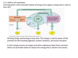
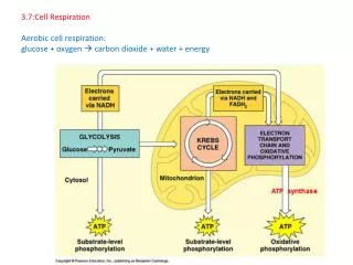
Essential Knowledge: 2.A.3 Organisms must exchange matter with the environment to grow, reproduce, and maintain organization2.D.2 Homeostatic mechanisms reflect both common ancestry and divergence due to adaptation in different environments.4.A.4 Organisms exhibit complex Properties due to interactions between constituent parts4.B.2 Cooperative interactions within organisms promote efficiency in the use of energy and matter
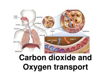
Circulation of Glucose, Oxygen, and Carbon Dioxide • How do simple organisms like jelly fish and flat worms exchange reactants and products of cellular respiration? • Simple animals have a body wall that is only two cells thick and that encloses a gastrovascular cavity • This cavity functions in both digestion and distribution of substances throughout the body • Materials can diffuse in or out of the gastrovascular cavity as needed.
Fig. 42-2 Circular canal Mouth Pharynx Mouth Radial canal 5 cm 2 mm (a) The moon jelly Aurelia, a cnidarian (b) The planarian Dugesia, a flatworm
Circulation of Glucose, Oxygen, and Carbon Dioxide • How do more complex organisms exchanges the reactants and products of cellular respiration? • They have either an open or closed circulatory system. • How does an open circulatory system work? • In insects, other arthropods, and most molluscs, blood bathes the organs directly in an open circulatory system • In an open circulatory system, there is no distinction between blood and interstitial fluid, and this general body fluid is more correctly called hemolymph
Circulation of Glucose, Oxygen, and Carbon Dioxide • How does a closed circulatory system work? • In a closed circulatory system, blood is confined to vessels and is distinct from the interstitial fluid • Closed systems are more efficient at transporting circulatory fluids to tissues and cells • What do we call our circulatory system? • Humans and other vertebrates have a closed circulatory system, often called the cardiovascular system • The three main types of blood vessels are arteries, veins, and capillaries
Fig. 42-3 Heart Heart Blood Hemolymph in sinuses surrounding organs Small branch vessels In each organ Interstitial fluid Pores Dorsal vessel (main heart) Tubular heart Auxiliary hearts Ventral vessels (a) An open circulatory system (b) A closed circulatory system
Circulation of Glucose, Oxygen, and Carbon Dioxide • What are the major components of the vertebrate circulatory system? • Arteries branch into arterioles and carry blood to capillaries • Networks of capillaries called capillary beds are the sites of chemical exchange between the blood and interstitial fluid • Venulesconverge into veins and return blood from capillaries to the heart • Vertebrate hearts contain two or more chambers • Blood enters through an atrium and is pumped out through a ventricle
Fig. 42-4 Gill capillaries Single circulatory loop with a 2 chambered heart Gill circulation Artery Ventricle Heart Atrium Systemic circulation Vein Systemic capillaries
Fig. 42-5 Double circulation with a 4 chambered heart Double heart circulation w/ 3 chambered Amphibians Reptiles (Except Birds) Mammals and Birds Lung and skin capillaries Lung capillaries Lung capillaries Right systemic aorta Pulmocutaneous circuit Pulmonary circuit Pulmonary circuit Atrium (A) Atrium (A) A A A A V V Ventricle (V) V V Left systemic aorta Left Right Left Right Right Left Systemic circuit Systemic circuit Systemic capillaries Systemic capillaries Systemic capillaries Double circulation with a 3 chambered heart – ventricle partially divided
Circulation of Glucose, Oxygen, and Carbon Dioxide • How does blood flow in a mammal? • Blood begins its flow with the right ventricle pumping blood to the lungs • In the lungs, the blood loads O2 and unloads CO2 • Oxygen-rich blood from the lungs enters the heart at the left atrium and is pumped through the aorta to the body tissues by the left ventricle • The aorta provides blood to the heart through the coronary arteries • Blood returns to the heart through the superior vena cava (blood from head, neck, and forelimbs) and inferior vena cava (blood from trunk and hind limbs) • The superior vena cava and inferior vena cava flow into the right atrium
Fig. 42-6 Capillaries of head and forelimbs Superior vena cava 7 Pulmonary artery Pulmonary artery Capillaries of right lung Aorta 9 Capillaries of left lung 3 3 2 4 11 Pulmonary vein Pulmonary vein 5 1 Right atrium Left atrium 10 Right ventricle Left ventricle Inferior vena cava Aorta Capillaries of abdominal organs and hind limbs 8
Fig. 42-7 Pulmonary artery Aorta Pulmonary artery Right atrium Left atrium Semilunar valve Semilunar valve Atrioventricular valve Atrioventricular valve Right ventricle Left ventricle
Circulation of Glucose, Oxygen, and Carbon Dioxide • How does the heart contract? What are the two phases of the cardiac cycle? • The contraction, or pumping, phase is called systole • The relaxation, or filling, phase is called diastole • What is another name for the heart rate? • The heart rate, also called the pulse, is the number of beats per minute
Fig. 42-8 Atrial systole; ventricular diastole 2 Semilunar valves closed 0.1 sec Semilunar valves open AV valves open 0.4 sec 0.3 sec 1 Atrial and ventricular diastole AV valves closed 3 Ventricular systole; atrial diastole
Circulation of Glucose, Oxygen, and Carbon Dioxide • What are the four valves in the heart called? And what is their purpose? • Four valves prevent backflow of blood in the heart • The atrioventricular (AV) valves separate each atrium and ventricle • The semilunar valves control blood flow to the aorta and the pulmonary artery • The “lub-dup” sound of a heart beat is caused by the recoil of blood against the AV valves (lub) then against the semilunar (dup) valves
Circulation of Glucose, Oxygen, and Carbon Dioxide • How does the heart maintain its rhythmic beat? • The sinoatrial (SA) node, or pacemaker, sets the rate and timing at which cardiac muscle cells contract • Impulses from the SA node travel to the atrioventricular (AV) node • At the AV node, the impulses are delayed and then travel to the Purkinje fibers that make the ventricles contract • The pacemaker (SA node) is influenced by nerves, hormones, body temperature, and exercise
Fig. 42-9-5 3 1 2 Pacemaker generates wave of signals to contract. Signals are delayed at AV node. Signals pass to heart apex. Signals spread throughout ventricles. 4 SA node (pacemaker) AV node Purkinje fibers Bundle branches Heart apex ECG
Circulation of Glucose, Oxygen, and Carbon Dioxide • How is the structure of blood vessels adapted to transport material throughout the body? • Capillaries have thin walls, the endothelium plus its basement membrane, to facilitate the exchange of materials • Arteries and veins have an endothelium, smooth muscle, and connective tissue • Arteries have thicker walls than veins to accommodate the high pressure of blood pumped from the heart • In the thinner-walled veins, blood flows back to the heart mainly as a result of muscle action, valves prevent back flow
Fig. 42-10 Artery Vein SEM Valve 100 µm Basal lamina Endothelium Endothelium Smooth muscle Smooth muscle Connective tissue Connective tissue Capillary Artery Vein Arteriole Venule 15 µm Red blood cell Capillary LM
Circulation of Glucose, Oxygen, and Carbon Dioxide • How does blood flow change as it moves from arteries to capillaries to veins? • Blood flow is fast in arteries due to pumping of the heart • Blood flow slows in capillaries as the volume from one artery spreads to feed an entire capillary bed – this is good it slows things down and allows for exchange of materials • Blood flow increases slightly in veins due to decreased surface area
Fig. 42-11 5,000 4,000 Area (cm2) 3,000 2,000 1,000 0 50 40 Velocity (cm/sec) 30 20 10 0 120 Systolic pressure 100 80 Pressure (mm Hg) 60 Diastolic pressure 40 20 0 Aorta Veins Arteries Venules Arterioles Capillaries Venae cavae
Circulation of Glucose, Oxygen, and Carbon Dioxide • How can blood flow through capillaries be controlled? • Two mechanisms regulate distribution of blood in capillary beds: • Contraction of the smooth muscle layer in the wall of an arteriole constricts the vessel • Precapillary sphincters control flow of blood between arterioles and venules
Fig. 42-15 Thoroughfare channel Precapillary sphincters Capillaries Arteriole Venule (a) Sphincters relaxed Arteriole Venule (b) Sphincters contracted
Circulation of Glucose, Oxygen, and Carbon Dioxide • Where does the critical exchange of nutrients and gasses takes place in the circulatory system? • The critical exchange of substances between the blood and interstitial fluid takes place across the thin endothelial walls of the capillaries • The difference between blood pressure and osmotic pressure drives fluids out of capillaries at the arteriole end and into capillaries at the venule end
Fig. 42-16 Body tissue INTERSTITIAL FLUID Capillary Net fluid movement out Net fluid movement in Direction of blood flow Blood pressure Inward flow Pressure Outward flow Osmotic pressure Arterial end of capillary Venous end