Download
1 / 45
470 likes | 560 Views
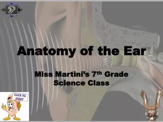
Learn the basic anatomic structures and physiology of the external, middle, and inner ear, as well as the functions and mechanisms of hearing and balance. Understand the skill objectives of ear examination.

E N D
ANATOMY AND PHYSIOLOGY OF THE EAR Yard.Doc.Dr.Müzeyyen Doğan
Learning goal and objectives of the lesson Learning goal of the lesson: The learner should know the basic anatomic structures and physiology of external, middle and inner ear Learning objectives of the lesson the learner will be able to: • identify the anatomic structures of the external, middle and inner ear • identify the physiology of the external, middle and inner ear • identify the physiology of the hearing • identify the physiology of the balance Skill objectives the learner will be able to ear examination
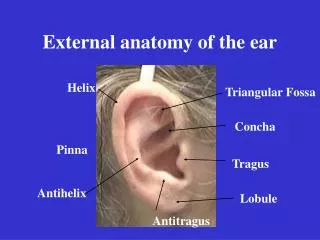
OUTER EAR • Consists of the auricle and EAM • Skin-lined apparatus • Approximately 2.5 cm in length • Ends at tympanic membrane
OUTER EAR • Auricle is mostly skin-lined cartilage • External auditory meatus • Cartilage: ~40% • Bony: ~60% • S-shaped • Narrowest portion at bony-cartilage junction
Anatomy and Physiology • EAC is related to various contiguous structures • Tympanic membrane • Mastoid • Glenoid fossa • Cranial fossa • Infratemporal fossa
Anatomy and Physiology • Squamous epithelium • Bony skin – 0.2mm • Cartilage skin • 0.5 to 1.0 mm • Apopilosebaceous unit
The Middle Ear: • A cleft within the temporal bone • Lining is mucous membrane • Tympanic Membrane separates it from EAC • Eustachian tube connects it to nasopharynx • Also Connected to Mastoid Air Cells
Middle Ear Structures 1- Malleus 2- Incus --Ossicles 3- Stapes 4- Tympanic Membrane (Eardrum) 5- Round Window 6- Eustachian Tube
Middle Ear Muscles 1.The StapediusAttaches to Stapes,Contracts in Response to Loud sounds, chewing, speaking; Facial (VIIth cranial) nerve 2. The Tensor TympaniHelps open Eustachian tube
Ligaments of Middle Ear Function restrict and confine the effect of ossicles to act as a lever restrict movements to reduce the chance of damage to the inner ear prevents distortion to sound
Middle Ear Functions • Impedance Matching • Filtering • Acoustic Reflex
Impedance Matching • The middle ear allows the impedance matching of sound traveling in air to acoustic waves traveling in a system of fluids and membranes in the inner ear
Acoustic Reflex • The movement of the ossicles may be stiffened by two muscles, the stapedius and tensor tympani, which are under the control of the facial nerve and trigeminal nerve, respectively. These muscles contract in response to loud sounds, thereby reducing the transmission of sound to the inner ear. This is called the acoustic reflex
MECHANISMS OF MIDDLE-EAR GAIN Acoustic Coupling Ossicular Coupling • Area Difference (TM to footplate) • Lever Action (Malleus to Incus)
Acoustic Coupling • The collected pressure of sound vibration that strikes the tympanic membrane is therefore concentrated down to this much smaller area of the footplate, increasing the force but reducing the velocity and displacement, and thereby coupling the acoustic energy.
Anatomy and Physiology • Ossicular Amplifier System
MIDDLE-EAR GAIN • The ossicles are classically supposed to mechanically convert the vibrations of the eardrum, into amplified pressure waves in the fluid of the cochlea (or inner ear) with a lever arm factor of 1.3. Since the area of the eardrum is about 17 fold larger than that of the oval window, the sound pressure is concentrated, leading to a pressure gain of at least 22.
Function of Middle Ear Conduction Conduct sound from the outer ear to the inner ear Protection Creates a barrier that protects the middle and inner areas from foreign objects Middle ear muscles may provide protection from loud sounds Transducer Converts acoustic energy to mechanical energy Converts mechanical energy to hydraulic energy Amplifier Transformer action of the middle ear only about 1/1000 of the acoustic energy in air would be transmitted to the inner-ear fluids (about 30 dB hearing loss)
INNER EAR INNER EAR : • Cochlea – hearing • Vestibule – static equilibrium • Semicircular canals – dynamic equilibrium
OHC vs. IHC Function • Vestibulocochlear nerve – cranial nerve VIII • Audible range: 20 -- 20,000 hertz • Ossicles amplify sound 22 X • Some nerve fibers cross over to opposite side of brain
Hair Cells Outer Hair Cells Inner Hair Cells OHC movie
OCH Cilia Theory: Tip-links <<<IHC OHC >>>
Equilibrium – Balance Static equilibrium – maintenance of body posture relative to gravity while the body is still. Dynamic equilibrium – maintenance of the body posture (mainly the head) in response to sudden movements. Tracking a moving object.
Static Equilibrium Inside the vestibule are two chambers : utricle and saccule. Regions of hair cells and supporting cells called maculae. Otoliths – “ear rocks”
Dynamic Equilibrium Semicircular canals In ampulla is the crista ampullaris – contains hair cells and supporting cells covered by a gelatinous mass called the cupula. Neurological connections between eyes and semicircular canals – for tracking Nystagmus