Download
1 / 15
150 likes | 174 Views
Explore the structures and functions of the outer, middle, and inner ear in hearing and equilibrium. Learn how sound travels through the ear and pathways to the brain. Interactive pages included.

E N D
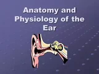
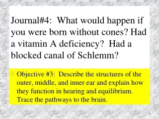
Journal#4: What would happen if you were born without cones? Had a vitamin A deficiency? Had a blocked canal of Schlemm? • Objective #3: Describe the structures of the outer, middle, and inner ear and explain how they function in hearing and equilibrium. Trace the pathways to the brain.
Main Components of the Hearing Mechanism: • Outer Ear • Middle Ear • Inner Ear • Central Auditory Nervous System
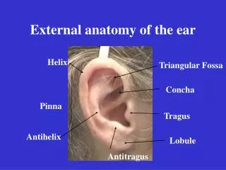
Structures of the Outer Ear • Auricle (Pinna) • Collects sound • Localization • Amplifies sound (approx. 5-6 dB)
External Auditory Canal: • Approx. 1 inch in length • “S” shaped • Lined with cerumen glands • Outer 1/3 surrounded by cartilage • Inner 2/3’s surrounded by mastoid bone
Mastoid Process • Bony ridge behind the auricle • Provides support to the external ear and posterior wall of the middle ear cavity
Tympanic Membrane: • Thin membrane • Forms boundary between outer and middle ear • Vibrates in response to sound • Changes acoustical energy into mechanical energy
The Ossicles: • A: Malleus • B: Incus • C: Stapes • Smallest bones in the body • Acts as a lever system • Footplate of stapes enters oval window of the cochlea • Stapedius Muscle • Connects stapes to wall of middle ear • Contracts in response to loud sounds (called the Acoustic Reflex)
Eustachian Tube (AKA: “The Equalizer”) • Lined with mucous membrane • Connects middle ear to nasopharynx • “Equalizes” air pressure
Structures of the Inner Ear • Cochlea • Snail shaped organ with a series of fluid-filled tunnels • Converts mechanical energy to electrical energy
The end organ of hearing Contains stereocilia and hair cells. Organ Of Corti:
Hair Cells: • Frequency specific • High pitches= base of cochlea • Low pitches= apex of cochlea
Vestibular System • Consists of three semi-circular canals • Shares fluid with the cochlea • Controls balance
Central Auditory System • VIIIth Cranial nerve or “Auditory Nerve” • Carries signals from cochlea to brain • Auditory Cortex • Temporal lobe of the brain where sound is perceived and analyzed
How Sound Travels Through The Ear... 1. Acoustic energy, in the form of sound waves, is channeled into the ear canal by the pinna 2. Sound waves hit the tympanic membrane and cause it to vibrate, like a drum, changing it into mechanical energy 3. The malleus, which is attached to the tympanic membrane, starts the ossicles into motion 4. The stapes moves in and out of the oval window of the cochlea creating a fluid motion 5. The fluid movement causes membranes in the Organ of Corti to shear against the hair cells 6. This creates an electrical signal which is sent up the Auditory Nerve to the brain The brain interprets it as sound!