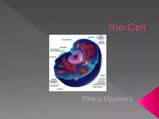
THE CELL
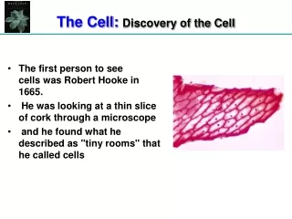
THE CELL. Cell - from the Latin word cella meaning small room. Robert Hooke (about 1670 ) - first to use term in a biological sense. Used it to describe the small, organized compartments he observed in dead cork tissue. Not empty - contained "jellylike" material - cell cytoplasm

THE CELL
E N D
Presentation Transcript
Cell - from the Latin word cella meaning small room Robert Hooke (about 1670) - first to use term in a biological sense. Used it to describe the small, organized compartmentshe observed in dead cork tissue Not empty - contained "jellylike" material - cell cytoplasm In animals the cell consists of plasmalemma (cell membrane) + cytoplasm containing various organelles and other inclusions CELL - may be defined as the smallest structural unit of a living organism that can function independently if placed in an appropriate environment. Cell Theory - developed independently by both Schleiden and Schwann in 1832. States that all living organisms are constructed of small sub-units called cells. 2
Composition of mammalian tissues 1. cells 2. intercellular substance 3. tissue fluid However, items 2 and 3 would not be present if the tissue did not contain cells. 3
So, where do the trillions of cells that compose your body come from? Egg + sperm -> Fertilization -> Zygote Cleavage + subsequent cell division Gastrulation - establishes primary germ layers ectoderm mesoderm endoderm Human egg Organogenesis - establishes organ primordia Histogenesis - establishes specific tissue types - this involves Cellular differentiation - specific function 4
HISTOGENESIS/CELLULAR DIFFERENTIATION -> MAJOR SPECIALIZATIONS OF CELLS 1. contraction - muscle cells 2. conduction of electrical impulses - nerve cells 3. storage of lipids - adipose cells 4. synthesis of enzymes and other substances - secretory cells 5. support of tissues, cushioning - connective tissue cells 6. protection - immune system cells, skin cells 7. gas transport - erythrocytes ETC. Cellular structure and function are linked! 5
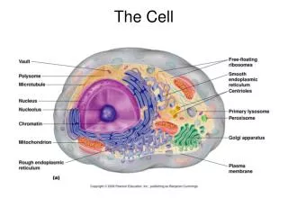
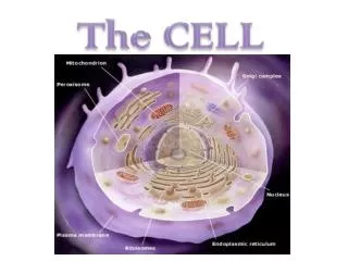
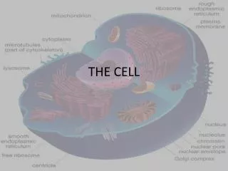
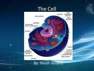
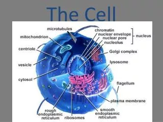
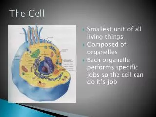
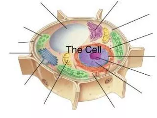
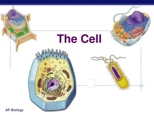
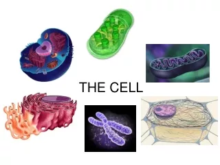
CELL STRUCTURE 3 major cell components 1. Plasmalemma = plasma membrane = cell membrane 2. Cytoplasm (fluid, soluble molecules, various organelles, other non-membranous inclusions) 3. Nucleus 6
THE PLASMALEMMA - CELL MEMBRANE Barrier between the cell and outside world. Determines what enters and leaves the cell. Classical model was a phospho-lipid bilayer with the hydrophilic phosphate radicals forming the outer surfaces and the hydrophobic fatty acid chains in between. http://www.cbc.umn.edu/~mwd/cell_www/chapter2/membrane.html 7
THE PLASMALEMMA - CELL MEMBRANE Fluid mosaic model - proteins embedded in or associated with lipid bilayer, some on one side or the other and others extending across bilayer. Most proteins that do not extend across are on inner surface adjacent to cytoplasm. Cholesterol is present in external surface. Proteins are not static - may move about in or on lipid bilayer. Movement mediated by cytoskeleton. Oligosaccharide components of glycoproteins extend from external surface forming the glycocalyx - functions in recognition and adhesion. 8
THE PLASMALEMMA - CELL MEMBRANE Molecular components of the plasmalemma: 1. phospholipids 9 http://www.jdaross.mcmail.com/cell2.htm
Molecular components of the plasmalemma: 2. proteins - integral (transmembrane) and peripheral (mostly on inner surface) 3. cholesterol and glycolipids - between some phospholipids in the external surface 4. glycocalyx - carbohydrate components of glycoproteins and glycolipids http://www.jdaross.mcmail.com/cell2.htm 10
CYTOPLASM Major constituents: 1. Cytoskeleton - microtubules and various filaments 2. Organelles - membrane bound structures 3. Cytoplasmic inclusions - non-membrane bound,non-filament structures, may be temporary. 11
Cytoskeleton Microtubules - 18 - 30 nm diameter: 1. Major skeletal component - “stiff” rods composed of 13 protofilaments - each 4-5 nm diameter 2. Function in support for cell structure, e.g. “hold-up” microvilli, pseudopodia - can be demonstrated with colchicine 3. May provide “skeletal” structure for contractile elements of cell to work against - necessary for movement 4. Provide pathways for transport of molecules, e.g. axonal transport 5. Form framework for flagella and cilia - 9+2 axoneme structure 6. Basis for mitotic spindle in conjunction with centrioles - important in movement of replicated chomosomes to opposite poles of mitotic apparatus. http://cellbio.utmb.edu/cellbio/microtub.htm http://www.physorg.com/news4689.html 12 http://www.rpi.edu/dept/bcbp/molbiochem/MBWeb/mb2/part1/kinesin.htm
Epithelial cell with labeled microtubules (red). Nucleus is yellow. Microtubule labeling - rhodamine conjugated to tubulin antibodies Nucleus - sytox green stain (DNA specific stain) 13 http://www.itg.uiuc.edu/technology/atlas/structures/nucleus/nucleus_07.htm
CYTOPLASMIC FILAMENTS Microfilaments - 5-7 nm diameter 1. Composed of actin 2. Responsible for cell movements e.g. bending of microvilli, contraction of pseudopodia, formation of cell division furrow 3. Actin is a component of sarcomere in striated muscle http://images.google.com/imgres?imgurl=http://www.arn.org/docs/glicksman/090104%2520fig2%2520actin.jpg&imgrefurl=http://www.arn.org/docs/glicksman/eyw_040901.htm&usg=__Wc8TIhLWWpddPqpUrbbhRzzvqV0=&h=389&w=480&sz=46&hl=en&start=11&um=1&tbnid=zYOy2Ggbp3yxPM:&tbnh=105&tbnw=129&prev=/images%3Fq%3Dsarcomere%2Bpicture%26hl%3Den%26client%3Dsafari%26rls%3Den%26sa%3DX%26um%3D1 14
Microfilaments - rhodamine conjugated phalloidin Nucleus - sytox green (DNA specific stain) 15 http://micro.magnet.fsu.edu/cells/microfilaments/microfilaments.html http://www.itg.uiuc.edu/technology/atlas/structures/ http://www.bio.miami.edu/~cmallery/150/cells/c8.6x26.microfilaments.jpg
Intermediate filaments- 10 - 15 nm diameter Comprise a variety of other filaments found in the cell, e.g. 1. Myosin filaments of sarcomere 2. Tonofilaments of desmosomes 3. Neurofilaments found in neurons 4. Components of terminal web at apical end of epithelial cells 16
Intermediate filaments - neurofilaments Confocal maximum intensity projection of guinea pig embryo labeled with neurofilament antibody visualized with a fluorescent probe. Labeling in brain, spinal cord, cranial nerves, and spinal nerves 17 http://microscopy.bio-rad.com/moviesandimages/Neuroscience.htm
CELLULAR JUNCTIONS 1. Zonula occludens 2. Zonula adherens 3. Desmosome structures a. macula adherens - desmosome b. hemidesmosome c. septate desmosome 4. Gap junction 5. Interdigitating membrane We will discuss these furtherwhen we talk about epithelia. 18
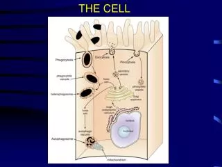
ORGANELLES The Mitochondrion (sgl.), Mitochondria (pl.) 1. Organelles specialized for the production of ATP 2. Recent cellular and molecular evidence -> leaves no doubt that mitochondria evolved from primitive bacteria that were symbionts of the ancestral precursor of the eukaryotic cell. 19 http://cellbio.utmb.edu/cellbio/mitoch2.htm http://micro.magnet.fsu.edu/cells/mitochondria/mitochondria.html
The Mitochondrion (sgl.), Mitochondria (pl.) 3. Length - 0.5 - 1.0 μm wide, up to 10 μm 4. Characterized by a double (inner and outer) membrane. The inner membrane forms folds called cristae. - intercristal space (matrix) between cristae. 5. Elementary particles on stalks that arise from cristae. 20 http://micro.magnet.fsu.edu/cells/mitochondria/mitochondria.html http://cellbio.utmb.edu/cellbio/mitochondria_1.htm#inner%20membrane
5. ATP production: • Glycolysis, which occurs in the cytoplasm outside the mitochondrion -- glucose converted to 2 pyruvate + 2 ATP • Pyruvate used to make acetyl co-enzyme A • In the mitochondrion acetyl co-enzymeA enters the Krebs cycle -> Krebs cycle reactions occur in matrix (intercristal space) -- results in free H+ and 2 ATP (one for each pyruvate molecule) • H+ processed through electron transport chain proteins on the cristal membranes and finally, the ATP synthase in the elementaryparticles gives rise to additional ATP. (oxidative phosphorylation) • Oxidative phosphorylation -> 30 - 36 ATP are produced for each molecule of glucose converted to carbon dioxide and water. 21
The Mitochondrion “Three-dimensional reconstruction of a dendritic mitochondrion from chick cerebellum. Created tomographically from a tilt series of a 0.5 micron section. This movie steps through every fourth slice of the tomogram, a three-dimensional image of the mitochondrion.” http://www.sci.sdsu.edu/TFrey/MitoMovie.htm 22
The Mitochondrion Another way of rendering tomographic data to reveal mitochondrial structure. Surface rendering. http://www.sci.sdsu.edu/TFrey/MitoMovie.htm 23
Endoplasmic Reticulum (ER) 1. A layered membrane bound organelle that consists of a network of tubular and vesicular cisternae that ramify throughout large volumes of cytoplasm. 2. Membrane of ER surrounds cavities that are within the ER. 3. Membrane of ER is continuous with outer nuclear membrane. 4. Site of processing of lipid and protein for cellular functions. 5. Rough ER -> ribosomes associated with outer surface -> processing of proteins 6. Smooth ER -> no ribosomes -> processing of lipids (e.g. steroids) 7. Sarcoplasmic reticulum - specialized smooth ER associated with striated muscle. Specialized for storage and release of Ca+. 24
The Golgi Body/Apparatus (called the dictyosome in plants) 1. An organelle consisting of interconnected stacks of flattened, membrane bound saccules that are called cisternae. 2. Plays role in packaging and processing of materials synthesized within cell, a. lysosomal enzymes b. secretory products 3. Convex side of stack -> forming (cis) face 4. Concave side of stack -> maturing (trans) face 25 http://library.thinkquest.org/C004535/golgi_apparatus.html
The Golgi Body 5. Materials synthesiszed and processed in ER -> carried in transfer vesicles to forming face -> vesicle contents emptied into cisternae -> further processing 6. Materials move through cisternae to maturing face -> packaged into vesicles that bud off maturing face. a. lysosomes b. secretory vesicles 7. These vesicles may fuse with each other in cytoplasm to form larger vesicles. 26
Visualizing the Golgi Apparatus 27 http://www.itg.uiuc.edu/technology/atlas/structures/
Electron tomogram - Golgi Apparatus 28 http://bio3d.colorado.edu/pubs/Golgi/Gset1.html
The Golgi Body/Apparatus Green fluorescent protein (GFP) was attached to a viral glycoprotein (VSVG) that moves through the secretory pathway to the cell surface. Time lapse movie shows the movement of this labeled protein (in transfer vesicles) from the endoplasmic reticulum to the golgi apparatus. 29 http://dir.nichd.nih.gov/cbmb/pb1labob.html
The cytoplasm is a crowded place. Tomographic 3-D reconstruction of a volume of cytoplasm “Figure 5. Stereo views of a model generated by tomographic reconstruction of a slab of cytoplasm from a cultured cell. The model displays the Golgi complex in the context of surrounding organelles: ER, yellow; membrane-bound ribosomes, blue; free ribosomes, orange; MTs, bright green; dense core vesicles, bright blue; clathrin-negative vesicles, white; clathrin-positive compartments and vesicles, bright red; clathrin-negative compartments and vesicles, purple; mitochondria, dark green. “ 30 http://www.jcb.org/cgi/content/full/153/6/F25
The Nucleus Largest and most conspicuous organelle of eukaryotic cells. Location of genetic material - DNA - chromosomes Site of DNA and RNA synthesis Molecular content (mainly the DNA) is basophilic - so nucleus takes up a dark/dense stain in many tissue preparations since stains are often basophilic At ultrastructural level - DNA is of two types in nucleus of non-dividing cell - unraveled, euchromatin, involved in synthetic activities. Condensed and obvious - heterochromatin DNA in dividing cell is highly condensed into typical chromosomes 31
General Nuclear Structure Nuclear pores allow molecules to get in and out of the nucleus, particularly mRNA, a very large molecule. 32
Nuclear pores The upper figure above shows a frontal view of the nuclear pore as though you are looking down on the pore. It contains 8 subunits that "clamp" over region of the inner and outer membrane where they join. Nuclear pores are formed at sites where the inner and outer membranes of the nuclear envelope are joined. Also, the heterochromatin, which carries the genetic material, is organized so that a space or "pathway" is created to the nuclear pore. This electron micrograph shown in the figure above depicts a transverse view of a nuclear pore complex seen with the transmission electron microscope. 33 http://cellbio.utmb.edu/cellbio/nuclear_envelope.htm#Pore
Nuclear pores In order to see the structure of the nuclear pore as diagramed in the preceding figure, special preparative techniques for electron microscopy have to be used. Freeze etching Negative staining How nuclear pores function is not well understood. 34
Nuclear pore complexes (NPCs) are embedded in the nuclear membrane and are about 9 nm in diameter. They regulate the movement of molecules and ions in and out of the nucleus. Small molecules and ions pass though the pores by passive diffusion. Large molecules like RNA and ribonuceloproteins move through the pores via active transport mechanisms that require ATP. To some extent movement through the pore appears to be regulated by opening and closing of the distal ring structure, an event that is caused by the presence or absence of calcium ions. Atomic force microscopy 35 http://www.scripps.edu/~stoffler/proj/NPC/npc.html
The Nucleolus Where ribosomal RNA is synthesized Composed of the nucleolar organizing center, pars granulosa, pars fibrosa, and a supporting matrix Nucleolar organizing center - location of rRNA genes Pars fibrosa = fibrillar zone surrounds the nucleolar organizing center, composed of newly formed rRNA Pars granulosa = granular zone outside pars fibrosa, composed of large and small ribosomal subunits. rRNA is synthesized from DNA in the nucleolus that is derived from a number of different chromosomes. The ribonucleoprotein components of ribosomes move out of the nucleus through the nuclear pores. They are assembled into ribosomes in the cytoplasm. Whole ribosomes cannot pass through the nuclear pores. 36
The Chromosomes Humans - 46 chromosomes, 23 pairs A fiber of chromatin consists of a single, long DNA double helix to which is added two kinds of proteins - histones and nonhistone chromosomal proteins. The chromatin fibers are dispersed within the nucleus, anchored at various places to the inner membrane of the nuclear envelope. http://www.csc.fi/jpr/emt/movies/chromosome.html http://life.nthu.edu.tw/~d867415/chromatin.html 37 http://www.people.virginia.edu/~js6s/zsfig/chromatin.html
Chromosomes Euchromatin - dispersed chromatin in nucleus of non-dividing cell - active in synthesis of mRNA Heterochromatin - condensed chromatin in nucleus of non-dividing cell 38
The inner life of a cell http://multimedia.mcb.harvard.edu/ 39
Cell division 1. Interphase - period between divisions, 3 major components, G1, S, G2. Synthesis of DNA takes place during the “S” stage. 2. Prophase - Replicated chromosomes condense (each chromatid is connected to its duplicate at the centromere), nuclear envelope breaks down near end of prophase, the nucleolus disappears, and the mitotic apparatus (mitotic spindle) forms. 3. Metaphase - Replicated chromosomes align on the “equator” of the mitotic apparatus. The chromotids are attached to the microtubules of the mitotic spindle at the centromere. 40
Cell division 4. Anaphase - Duplicate chromatids separate at the centromere and move toward opposite poles of the mitotic spindle. 5. Telophase - Chromosomes reach the poles of the mitotic spindle, nuclear envelope (membrane) reforms, cell completes division. 41
Cytoplasmic Inclusions 1. Lipid droplets 2. Glycogen 3. Melanin (pigment) 4. Lipofuscin (pigment) 5. Crystals of various types 6. Hemosiderin (pigment) 43
Lipid droplets http://www.medinfo.ufl.edu/year1/histo/images/d14a.jpg 44
Glycogen granules “Ewing's sarcoma of bone with extensive cytoplasmic glycogen deposits, electron micrograph.” 45 http://www.medlib.med.utah.edu/WebPath/HISTHTML/EM/EM027.html
Lipofuscin “Easily seen at the poles of the cardiac nuclei, brown-red in color. ... As one ages, the amount of lipofuscin will continue to increase, especially in the heart. One can determine whether a heart is relatively young or old, as a direct reflection of the amount of lipofuscin present.” http://erl.pathology.iupui.edu/C603/LABEL49.HTM 46 http://uhsweb.edu/tdemark/0052.htm
Hemosiderin “The brown coarsely granular material in macrophages in this alveolus is hemosiderin (iron oxide) that has accumulated as a result of the breakdown of RBC's and release of the iron in hemoglobin. The macrophages clear up this debris, which is eventually recycled.” 47 http://www-medlib.med.utah.edu/WebPath/CINJHTML/CINJ041.html