Download
1 / 41
410 likes | 553 Views
The Medical Defense in Mesothelioma Cases. Edward M. Slaughter Hawkins Parnell Thackston & Young LLP 4514 Cole Avenue Dallas, TX 75205. Overview. Mesothelioma: The Basics Stains Malignancy Tissue Digestion. MESOTHELIOMA: THE BASICS. Pleural

E N D
The Medical Defense in Mesothelioma Cases Edward M. Slaughter Hawkins Parnell Thackston & Young LLP 4514 Cole Avenue Dallas, TX 75205
Overview • Mesothelioma: The Basics • Stains • Malignancy • Tissue Digestion
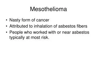
MESOTHELIOMA: THE BASICS • Pleural • Usually caused by asbestos or some similar fiber, but not always • Peritoneal • Maybe caused by asbestos 50% or less • Pericardial • Rarely caused by asbestos • Testicular mesothelioma • Too uncomfortable to discuss
Pleural Mesothelioma • Caused by prior radiation therapy at the site • Other fibers • Erionite (which is not just in Turkey) • Taconite • Maybe any fiber that that is biopersistent? • Spontaneous or ideopathic
Peritoneal Mesothelioma • Often caused by some other inflammatory process • Be on the lookout for: • Prior hernias • Diverticulitis • Hysterectomies • Anything that leads to chronic inflammation
Pericardial and Testicular • If you ever see one we can talk about then.
Types of mesothelioma • Epithelial • Sarcomatoid • Biphasic
Extremely Rare Diagnosis • Desmoplastic • Really just a rare subtype of sarcomatoid • Hard to diagnose and easily confused with sarcomatoid adenocarcinoma • Benign mesothelial hyperplasia • Reactive process that looks like malignant mesothelioma • Well differentiated papillary mesothelioma • benign
Old men get Mesothelioma • Female mesotheliomas are more often from another cause – maybe 50-50 • Really young people usually don’t have sufficient latency • If the plaintiff is young look for other causes
Take Away #1 – Be Suspicious • Mesothelioma in any location other than the pleura (by the lung) is very suspect • Mesothelioma diagnosis with extra words is suspect (wdpm, hyperplasia, etc) • Mesothelioma in young people and women is suspect. • Be suspicious of the diagnosis in these cases.
Stain Limitations • Can’t show if process is malignant or benign • No single stain can definitively diagnose mesothelioma • Glut-1 Caveat
Specificity v. Sensitivity EXAMPLE: LeuM1 STAIN • LeuM1 Stain Specific to Adenocarcinoma • LeuM1 Stain Does Not Have High Sensitivity though, because does not react with all adenocarcinoma
Other Common Stains • Cytokeratins • Glycoproteins • Calretinin • Thrombomodulin • HBME-1 • Cadherins • TTF-1
Take Away #2 – Stains are just a tool • IHC stains are NEVER enough for a complete diagnosis • They are positive stains and marginally positive stains • No one stain is enough • Stains cannot distinguish between malignant and benign mesotheliomas
Diagnosing Malignancy • Invasion • Necrosis • Clinical Correlation • Other factors
Stromal Invasion “True stromal invasion is by far the most reliable criterion of mesothelial malignancy” Andrew Churg M.D., et al The Separation of Benign and Malignant Mesothelial Proliferations; The American Journal of Surgical Pathology 24(9):1183
Minimal Invasion 06/29/05 - Report of Dr. J.F. Legier …One single slide, 589D4, shows early invasive disease with minimal invasion of fibrous stroma by mesothelial clusters.
6 Days Earlier – Same Pathology 06/23/05 - Report of Dr. J.C. Maddox …based on the clinical history and imaging studies that showed progression of the disease (“malignant mesothelioma on the left with extensive mediastinal invasion…”) … the patient more likely than not had a malignant mesothelioma that caused his death.
And Another Opinion 05/21/07 - Report of I. Allen Feingold Despite all of the above hisotpathological and even immunohistochemical evidence against malignancy in this case, it is important to consider the observation of Dr. Robert Viggiano who saw the patientin consultation and follow up…
Only Seen 10 Times Q: Okay. And out of those 7- or 800 cases, how many times have you had a difficult diagnosis like this, where you had to compare this sort of a benign, reactive process to a malignant mesothelioma? A: Maybe ten times. Dr. Jacques Legier, pp. 72-73
Clinical Correlation • Just fancy talk for “Did the patient seem like a man with mesothelioma?”
Necrosis • Is there dead or dying tissue?
Other Factors • Read this: Andrew Churg M.D., et al The Separation of Benign and Malignant Mesothelial Proliferations; The American Journal of Surgical Pathology 24(9):1183
Take away #3 – Not all Mesotheliomas are Malignant • Stains don’t prove malignancy • Invasion, Necrosis, Clinical Correlation do
Tissue Selection • A whole lung is ideal, but not necessary • Autopsy, Pneumonectomy or biopsy • Uninvolved tissue (not the tumor) • The Bigger, The Better • Multiple Sites • But as little as a gram can be enough
Results of Fiber Burden Analysis • Evidence of Fiber Type • Amount of Exposure • Evidence of Other Exposures • Helpful with Alternative Exposure Defenses
Take Away # 4 Digestion can be valuable • To prove amphibole exposure • To suggest an absence of chrysotile • To prove a dose estimate
All 4 Take Aways orThe Big Finale • Be Suspicious of unusual mesotheliomas (extra words, people under 60, women) • Stains are imperfect and don’t prove malignancy - just differentiate between mesothelioma and other processes • Malignancy has to be proven – invasion, necrosis, clinical correlation • Digestion can prove fiber type and dose, but not diagnosis