Download

1 / 48
480 likes | 628 Views
An Analysis on Cytology Diagnoses of the Effusion using LBC Technology. Jong Yull Kim, Prof.,PMIAC Eulji University Cellntech Bio.,Co.,Research Institute Zhanar Yeleubayeva, MD.,MIAC. 1. Kinds of Effusion using LBC Technology. Pleural fluid Ascitic fl ui d Cardiac fluid

E N D
An Analysis on Cytology Diagnoses of the Effusion using LBC Technology Jong Yull Kim, Prof.,PMIAC Eulji University Cellntech Bio.,Co.,Research Institute Zhanar Yeleubayeva, MD.,MIAC 1
Kinds of Effusion using LBC Technology • Pleural fluid • Ascitic fluid • Cardiac fluid • Synovial fluid 2
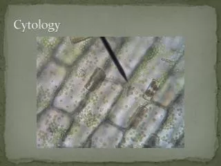
Specimen shows varied and different kinds of features of solutions 3
Conventional methods are considered as difficult to effectively smear -Bloody effusion -Mucous effusion -Clear effusion 4
Conventional methods are sometimes causing false negative and false positive. Bloody smear Dirty background & overlaping Degeneration 5
Main purpose of LBC technology -Thin smear -Clean background -Less degeneration than conventional method • use specially-designated equipment for smear. specially-designated filtering membrane. specially-designated reagent solution. 6
Process for specimen of effusion • Step 1. Sampling from patient by physician • Step 2. Transfer to cytopathologic laboratory -Effusion derived from patients should immediately be transferred to cytology rooms to prevent cellular degeneration -Cellular degeneration starts 30 minutes after sampling of effusion -More delay means increased intensity of degeneration -We should prepare the sample immediately to block cellular degeneration in labs 7
Step 3.Classify specimen according to feature of effusion • for pre-treatment 8
Step 4. Treatment ofspecimen according to feature of effusion • In case of non-bloody effusion & clear effusion • (generally common method : non-treatment of background) - after centrifuge at 1500~2500 rpm / 5 mins • - move sediment to preserve solution • - smear cells on slide by divice • - staining 9
2. In case of bloody effusion - centrifuge • - add “hemolysin” solution for hemolysis • - centrifuge • - move sediment to preserve solution • - smear cells on slide by divice - staining 10
3. In case of mucous effusion - centrifuge - add “mucosin” solution for discarding mucus - moving sediment to preserve solution - smearing cells on slide by device - pap staining 11
Increasing diagnostic rate Percentage of increasing diagnostic rate • Percentage of increasing • diagnostic rate • Among the Inadequate specimen Inadequate diagnosis 100 con con con LBC LBC LBC Percentage 80 60 40 20 More than mean 22% of diagnoses among the each inadequate specimen are diagnosed to sufficient specimen as either benign or malignant. 10 24% 19% 21% Blood Mucus Clear Source: Cellnatech bio, research institute. 12
In conclusion • Slide smear using the LBC technologies have advantages compared to two conventional methods. -The strengths follow: Eliminating backgrounds around diagnostic cells effectively makes easy microscopic work. -As LBC technology enables to manufacture thin smear, a deep and thorough reviews of morphological features in diagnostic cells are guaranteed. -As the LBC technology enables to allow samples to have proper substance of background, identification for details of malignant cells like mucus producing adenocarcinoma and signet ring cell type adenocarcinoma is useful. • Based on different status of specimen, -the LBC technology could produce a series of slides. Meanwhile, re-examination and special stain are also posible. -As diagnostic cells exit in a slide by forming groups, it's also possible to find the origin of malignant details in metastasis. 13
Diagnosis 14
Body Cavity: potential space lined by mesothelium • Mesothelium : Single layer of mesothelial cells • Supporting vascular connective tissue 15
Condition with Neutrophilsin effusion (Abscess) dirty background with neutrophils clean background with neutrophils 16
Condition with RBC In Effusion (Traumatic hemorrhage) bloody background with lymphocytes clean background with lymphocytes 17
Condition with Eosinophilsin Effusion (parasitic infestation) portein background with Eosinophils clean background with Eosinophils 18
Parasitic Infestation Parasitic Ova with Inflammatory and necrotic debris Parasitic Ova with clean background 19
Tuberculous Effusion • (Caseous necrosis) amorphous necrotic materials with lymphoid small particle. “Caseous necrosis” 20
Lymphocytes & epithelioid cells Low power feature 21
Rheumatoid Arthritis • Elongated histiocytes(carrot cells) Macrophages Amorphous proteinaceous debris 23
Multinucleated giant cells(MGH) Abundant clumps of granular debris • Monolobular or polylobular neutrophils • (degenerating neutrophils) 24
Systemic Lupus Erythematosus • LE cells ; enlarged neutrophil nucleus + pink to purplishamorphous • round intracytoplasmic mass • Degenerating cells(atypical plasmacytoid cells) • Nuclear debris 25
Mesothelial Reaction dirty background and degenerative change of mesothelial cells clean background and well preserved chromatin pattern. 26
Malignant mesothelioma & Mesothelial reaction many mesothelial cells with thick cytoplasm We can find different morphology of nuclear between both slides 27
Pseudomyxoma peritonei Viscous and thick mucinous material • Well-differentiated columnar and over distended by large mucinous • containing vacuoles • Benign looking columnar cells 28
Serous cystadenocarcinoma of ovary clean background and well preserved malignant cluster dirty background and degenerated malignant cluster Papillary or rosette formation Often intracytoplasmic large vacuoles 29
Muciouscystadenocarcinoma of Ovary Gray to deep purple colored mucus & abundant, well defined cytoplasm • Irregular shape in nuclear border with coarse chromatin,large & bizarre nucleoli 30
Signetic ring cell type adenocarcinoma from stomach • Cellular group • Glandularstructure • Indibidual pattern • Similar histiocyte • Eccentric nucleus • Macronucleoli • Individual cells mucicarmine Stain positive intracytoplasmic mucus in signet ring cell carcinoma. Pap stain. Macronucleoi & eccentric nucleus 31
Signetic ring cell type adenocarcinoma from stomach dirty background with degenerated cancer cells clean background with well preserved cancer cells 32
Metastatic Adenocarcinoma From colon Tall columnar, multi-layer, tubular formation Necrotic tumor cells with necrotic background 33
Adenoid cystic carcinoma Refractive spherules have three-dimensional cancer cells with benign looking. 34
Giemsa stain pattern as intracystic pinkish materials Pap stain pattern as refractive three-dimensional appearance 35
Ductal carcinoma, metastatic structure of ball form in Ductal carcinoma, metastatic cannibalism in Ductal carcinoma, metastatic. 36
Mucinous carcinoma from Breast small amount of mucus background so much mucus background 37
Breast-Lobular carcinoma String of small mlignant cells with internuclear space. We called it as “Indian file appearance”. 38
Lobular ca. vs. Small cell ca. indian file appearance with scanty or abscentinternclear spaces indian file appearance with internclear spaces. 39
Small cell carcinoma Pap stain pattern including nuclear moulding & scanty cytoplasm Giemsa stain pattern including nuclear moulding & scanty cytoplasm 40
Small cell carcinoma, metastatic clustes of small cell carcinoma, metastatic. compact & tight clusters with scanty cytoplasm are remarkable. 41
Large cell carcinoma, metastatic marked nuclear pleomorphism & nuclear nippling in large cytoplasm. 42
Squamouscell carcinma, metastatic well preserved cancer cells with small amount of necrosis necrotic background with degenerated cancer cells 43
Lymphocytes vs. Malignant lymphoma(Body fluid) mature lymphocytes including some pseudofiber cells of lymphoma including open chromatin & nucleoli 44
Hodgkin’s lymphoma Reed-Sternberg cell Single prominent nucleoli • Variable amounts of immature lymphocytes • and plasma cells in background 45
Plasmocytoma Dysplastic plasma cell(myelocytes) with accentric nucleus including macronucleoli in Pap Dysplastic plasma cell(myelocytes) with accentric nucleus including intracytoplasmic clear zone in Giemsa 46
Malignant melanoma Malignant melanoma • Singly scattered or occa. In groups • Finely granular brown or black melanin pigments • Round-oval nuclei, central or eccentric nuclei • Prominent nucleoli, often bizarre forms 47