Download

1 / 28
280 likes | 406 Views
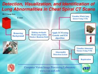
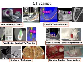
Detection, Visualization, and Identification of Lung Abnormalities in Chest Spiral CT Scans. 3D CT Image Data. Visualize Whole lung tissues Using VTK. 8 mm. Making stochastic Model using Gibbs Markov Random Field. Apply ICM using Genetic and EM algorithm. Removing Background.

E N D
Detection, Visualization, and Identification of Lung Abnormalities in Chest Spiral CT Scans 3D CT Image Data • Visualize Wholelungtissues Using VTK 8 mm • Making stochastic Model using Gibbs Markov Random Field • Apply ICM using Genetic and EM algorithm Removing Background • Visualize Abnormal Tissues Using VTK • Abnormality Detection System Registration Computer Vision Image Processing Laboratory www.cvip.uofl.edu
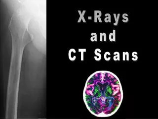
Medical Imaging Types of medical Imaging 1. X-ray Imaging Advantage Cheap Disadvantage It is just a projection of an object Computer Vision Image Processing Laboratory www.cvip.uofl.edu
Example of X-ray Imaging Computer Vision Image Processing Laboratory www.cvip.uofl.edu
Example of X-ray Imaging Computer Vision Image Processing Laboratory www.cvip.uofl.edu
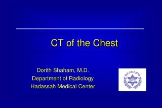
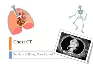
2. computed tomography (CT) • Advantage • better Geometry of the scanned subject • Using CT we can build 3-D model of the scanned subject • 3. Give high contrast between bones and soft tissues Computer Vision Image Processing Laboratory www.cvip.uofl.edu
Disadvantage 1. Ct has harmful effect due to radiation dose (X-ray) Computer Vision Image Processing Laboratory www.cvip.uofl.edu
Example of CT Computer Vision Image Processing Laboratory www.cvip.uofl.edu
3. Magnetic Resonance Imaging (MRI) Advantage 1. Give high contrast of soft tissues Disadvantages 1. Does not preserve the geometry of the scanned subject if it is compared with CT Computer Vision Image Processing Laboratory www.cvip.uofl.edu
Example of MRI Computer Vision Image Processing Laboratory www.cvip.uofl.edu
4. Ultrasound Imaging • Advantage • Real Time Imaging • No harmful effect Computer Vision Image Processing Laboratory www.cvip.uofl.edu
Example of Ultrasound Imaging Computer Vision Image Processing Laboratory www.cvip.uofl.edu
Abnormality Detection System Automated Lung Abnormality Detection System • Visualize Wholelungtissues Using VTK 3D CT Image Data 8 mm • Making stochastic Model using Gibbs Markov Random Field • Apply ICM using Genetic and EM algorithm Removing Background • Visualize Abnormal Tissues Using VTK Registration Computer Vision Image Processing Laboratory www.cvip.uofl.edu
System Design • Preprocessing Data • Such as you can filter your images in order to reduce the noise • LPF 2. HPF 3. BPF • 3. Median filter 4. Gaussian Filter Computer Vision Image Processing Laboratory www.cvip.uofl.edu
Image 3 x 3 pixel Computer Vision Image Processing Laboratory www.cvip.uofl.edu
Original imageImage after removing background 1. Remove the background Starting from the edge of the image, neighboring pixels are compared. Pixels having the same gray levels are removed (I.e., belong to the same region), while those differing are kept. Original Image 3x3 pixels Image 3 x 3 pixels after applying the algorithm Computer Vision Image Processing Laboratory www.cvip.uofl.edu
Background Chest Lung Computer Vision Image Processing Laboratory www.cvip.uofl.edu
How To estimate the Initial Mean for Lung and Chest? Computer Vision Image Processing Laboratory www.cvip.uofl.edu
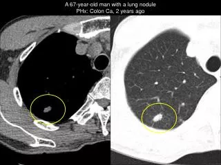
Abnormal tissues CT Slice Contain Abnormal Tissues Computer Vision Image Processing Laboratory www.cvip.uofl.edu
Slice_No. 32 Slice_No. 33 Abnormal tissues Computer Vision Image Processing Laboratory www.cvip.uofl.edu
Abnormality Detection Criteria • Each Ring Shape will take three ranks • Radial uniformity (R) • Position of the ring shape relative to the center of right or left lung edge (P) • Connectivity between different slices (C) Computer Vision Image Processing Laboratory www.cvip.uofl.edu
Abnormality System detection Abnormal Tissues yes Compute The Total Rank (R) for Each ring shape Detecting ringshape Remove the Normal Tissues R> 2 No Normal Tissues Computer Vision Image Processing Laboratory www.cvip.uofl.edu
a. Removing the normal tissues In order to remove the normal tissues of the lung, we will compute the histogram for each slice and search for its peak, and then remove all pixels beneath this peak. Before Removing normal Tissues After Removing normal Tissues Histogram of the CT slice Computer Vision Image Processing Laboratory www.cvip.uofl.edu
c. Ranking • NR, measures the uniformity distribution of the edges. • NC, measures the connectivity that the pixel (x, y) appears in the same location in different slices • NP, each pixel given a rank NP reflecting its position relative to the center of the right lung or the left lung. Total Rank (N)= NR + NC + NP Computer Vision Image Processing Laboratory www.cvip.uofl.edu
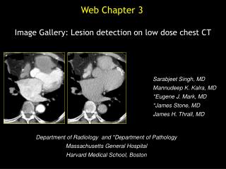
4. Results (a) Original slice from a spiral CT scan of a patient (b) Slice after removing the background (c) Desired tissues (e) The isolated lungs Computer Vision Image Processing Laboratory www.cvip.uofl.edu
(f) Bronchi, bronchioles and abnormal tissues (h) Manual detection by expert doctor (g) Abnormal tissues detected by our algorithm Computer Vision Image Processing Laboratory www.cvip.uofl.edu
Building 3-D model We use VTK tool to build 3-D model for the whole lung tissues and abnormal tissues, bronchi, and bronchioles 3-D model for the whole lung tissues Computer Vision Image Processing Laboratory www.cvip.uofl.edu
This Figure shows the abnormal tissues in the 3-D Computer Vision Image Processing Laboratory www.cvip.uofl.edu
More Results Computer Vision Image Processing Laboratory www.cvip.uofl.edu