Download
1 / 27
1.95k likes | 6.98k Views
Chest CT. By: Sara al-lithey, Nora Alanazi. Outline :. Chest CT. Indications. Contraindications. Chest CT protocols. - HRCT protocol. - Pathology protocol. - Pulmonary Embolism. Patient after care.

E N D
Chest CT By: Sara al-lithey, Nora Alanazi
Outline: • Chest CT. • Indications. • Contraindications. • Chest CT protocols. - HRCT protocol. - Pathology protocol. - Pulmonary Embolism. • Patient after care .
Chest CT scanning • is a noninvasive medical test that helps physicians diagnose and treat medical conditions. • CT scan is better for chest • CT it show many different types of tissue, including the lungs, heart, bones, soft tissues, muscle and blood vessels in short time.
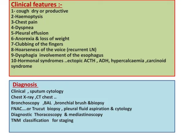
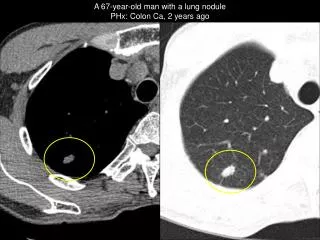
Indication • Chest pain. • Chest trauma. • Infection. • Intra-thoracic bleeding • Pulmonary embolism. • Pneumonia. • Suspected tumor or mass. • Assessment of interstitial lung disease • Minor fibrosis
Contraindication • Pregnancy. • Hypersensitivity to iodinated contrast media( if contrast is used). • Renal failure. • Heart disorder
1- HRCT • Without contrast. • High kV, mAs & thin slices to produces a high spatial resolution and anatomic detail • No preparation • Sedation if needed.
Patient position • Supine, in the center of the table • Feet first in the gantry (child, head first to see the chest movement during the breathing after the sedation). • Table height • The arms are above the head. • The external laser liner in the chine. Scout Images: • PA : plane 180º • Lat : plane 90º
Scanparameters: Window
Reformatting: sagittal & coronal Note: FOV is as small as possible while still including all of the soft tissue and the upper abdominal part (to check the metastasis).
2- Pathology (C+) Preparation: • NPO 3-4 hours before the procedure. • Not allergic or asthmatic. • Renal function test normal 1 week inpatient. 3 month diabetic patient 6 month non diabetic patient. • Canulla in the Rt arm, size 18, 20 Gag. • Sedation if needed
Procedure • Inspiration technique: As HRCT • Patient position: As HRCT • IV Contrast media: • Omnipaque • Xenetix • Adult : 300 • Child: 250
Contrast media The injector machine Hand injection Adult Child FR 5ml/sec 5ml/sec V 100 ml Weight × 2 Time delay 15- 20 sec C. in the Aorta
Scan parameters & Window : as Inspiration HRCT • 2nd Reconstruction: 1.25 ×1.25 • Axial & Sagittal – Soft window
3- Pulmonary Embolism scan • Fast helical CT scanning to visualization of the pulmonary vessels and lung parenchyma during the injection of a CM to detect any obstruction. • Optimal contrast should be in the pulmonary arteries, but not in the pulmonary veins. • Patient Preparation & position:As 2nd tech.
Main thing: calculate the contrast time that start from the injection until it will enter in the main pulmonary artery Two ways to calculate the timing: Smart Prep Bolus timing Then selected in the computer.
A- Smart Prep • In case if: • The heart rate abnormal (above 110-140) • Obese patient (above 110 Kg).
Scan parameters Window
b- Bolus Timing • In case if the heart rate normal (70-90) • Procedure • PA scout (to localize the pulmonary trunk). • FOV only in the bifurcation of the trachea and take CT images.
Select 20 images in the same location • Give 20 ml IV CM (FR 5 ml/ sec) • Start the CM in the same time of the exposure • wait the image to review. • From the 20 images, select the good image with the good contrast in the Main Pulmonary artery. • Time delayed calculation: • image number × 2 = sec
Then do the normal image from down- up with same before and fixed the scan time manually. • This graph shows that the contrast injected into the vein reached the pulmonary trunk in 8 seconds.
I.V. Contrast • Smart Prep: Adult: 100 ml – 5 ml/ sec • Bolus timing: Scan time × 5 = volume The bolus timing is Better than the Smart Prep • Less C. dose, so there is chance to repeat the scan again if there is any mistake ( the max dose is 100 ml/ day).
Reformatting • Sagittal • Coronal • Both oblique • Maximal intensity projection (MIP)
After care • Pt. can eat or drink as normal. • Drink fluids to flush the contrast. • Bandaged contrast injection site. • Watch the patient for adverse contrast reactions.