Download
1 / 29
290 likes | 704 Views
Transport of Solutes Across Plasma Membrane (II). Facilitated Transport Passive Facilitated Transport Active. Introduction.

E N D
Transport of Solutes Across Plasma Membrane (II) Facilitated Transport Passive Facilitated Transport Active
Introduction • In the last lecture, we studied diffusion and osmosis. This PPT is on facilitated transport of solutes. The term facilitated means “aided by” or “made possible by” proteins in the plasma membrane. These proteins will function as carriers, channels or pumps. • Study these slides and read relevant pages in Guyton. • In the lecture we’ll compare and contrast diffusion with facilitated transport and look at their significance in the context of physiological processes like absorption of nutrients in the gastrointestinal tract. • There are 28 slides
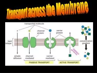
How Solutes Cross Membranes • Solutes move across cell membranes by: • Simple diffusion: We studied it last week • Facilitated transport: If they are relatively large, polar or charged (see next slide for examples). Facilitated transport may be: • Passive transport (this is also called facilitated diffusion) OR • Active (this is also called active transport) • Very large solutes (like proteins) move across the plasma membrane by bulk transport, called: • Endocytosis: For transport into the cell OR • Exocytosis: For transport out of the cell
Relative Permeability of Synthetic Bilayers to Some Solutes (Alberts Fig. 12.2)
Facilitated Transport • Facilitated transport may be characterized as • Active if it is endergonic OR • Passive, if it is exergonic. (Passive facilitated transport is also called facilitated diffusion) • It is mediated by membrane proteins and it is for solutes that are: • large and polar (previous slide for examples) • for cations and anions (previous slide for examples)
Types of Transport Proteins • The proteins that mediate facilitated transport are called: • Transporters • Also called carriers, pumps, permeases • Mediate either active or passive facilitated transport • Channel proteins • Ion channels, porins, aquaporins • Channel proteins mediate passive transport, always • They are all multipass proteins
Characteristics of Transporters • Proteins that function as transporters • Are allosteric • Have binding sites for one or more solutes • May behave as: • Uniports or • Coupled* transporters, and coupled transporters may function as: • symports (also called symporters) or antiports (also called antiporters) • Are solute specific • Exhibit Michaelis-menten kinetics * • Specificity, Vmax, Km, • Inhibition (competitive & noncompetitive)
Characteristics of Channels • Some are allosteric some are not allosteric • They are very selective for specific ions • Exist as two types, called • Leak channels (always open) Or • Gated channels. Gated channels fluctuate between: • open <-> close <-> inactivated states • Types of Gated channels • Voltage-gated: Open/close in response to changes in Vm • Ligand-gated: Open/close in response to ligand binding to receptor • Mechanosensitive: Open/close in response to forces (pressure, tension…)
Facilitated Transport ofNon-Charged Solute • Passive transport of non-charged solutes is • Mediated by transporters • Driven by magnitude of gradient (S) • Down gradient and toward equilibrium • Net flow is in either direction (into or out) • Exhibits Michalis-Menten kinetics
Facilitated Transport of Ions • Facilitated transport of ions is mediated by channels- leak or gated • Is down gradient and towards equilibrium • Is driven by electrochemical gradient • The electrochemical gradient takes into account both the membrane potential plus the concentration gradient • Depending on the charge of the ion, the membrane potential may favor or oppose influx or effluxof the ion
Effect of Electrochemical Gradient on Ion Flux (Study legend Fig. 12.8)
Active TransportStudy Guyton Ch. 4 Carried out by Coupled Transporters ATP-Driven Pumps Light-Driven Pump (not in human cells)
Active Transport(1): Significance • Active transport is important for: • Intake of nutrients and solutes from the extra-cellular fluid even when the concentration of these solutes is higher inside the cell. • For moving wastes or excess ions out of the cell even their concentrations is higher in the extracellular fluid. • For maintaining non-equilibrium concentrations of certain ions across the plasma membrane (or across membrane of certain organelles • Last item is essential for sustaining life. Many cellular functions (like nerve impulse conduction) depend on these concentration gradients. • Active transport may be classified as: • 1) primary or direct if coupled to ATP hydrolysis • 2) secondary or indirect if not directly coupled to ATP hydrolysis. This type is coupled to the potential energy in a Na+ or H+ gradient
Active Transport (2): Some facts • Mediated by uniports, symports or antiports • Kinetics more complex than Michalis’ • Against gradient and away from equilibrium • Is inherently unidirectional or vectorial • Is energy - dependent • May be classified as: • primary or direct • Secondary or indirect
Indirect Active Transport • Uses potential energy in Na+ or H+ concentration gradient • Na+ in animals • H+ in plants, bacteria and mitochondria • One solute is transported down its gradient concomitantly (together) with another solute transported against its gradient
Direct Active Transport • Direct active transport is coupled to: • ATP hydrolysis or to • Light energy (in some prokaryotes) • Direct active transport depends on four types of transport ATP’ases described in the next six slides
Types of Proteins involved in Primary Active Transport • The proteins involved in active transport are classified as: • Tranport ATP’ases or ATP-driven pumps (Na+/K+ pump) • Light-driven pumps (like bacteriorhodopsin) • Coupled transporters • There are four types of transport ATP-ases • P-Type (P stands for phosphate group) • V-Type (V stands for vacuoles, vessicles) • F-Type (F stands for factor) • ABC-Type (ATP Binding Cassette-Type) • Relevant details are given in the next 4 slides
P-type ATPases • Example: The Na+/K+pump • They are found in the plasma membrane of most animal cells, plants and fungi and in the sarcoplasmic reticulum of muscle cells. • They are reversibly phosphorylated by ATP. Phosphorylation-dephosphorylation is an intrinsic event in the transport process. • All of them transport cations (Ca2+, H+, Na+ and K+) • Sensitive to inhibition by the vanadate ion (VO4)3-
V-type ATPases • Found mostly in the membrane of plant vacuoles and in that of lysosomes. • They pump H+ ions and help maintain a proton gradient that ranges between 10x to 10,000x • They consist of two multimeric subunits, an integral and a peripheral one. Only the peripheral component (it faces the extracellular fluid) gets phosphorylated. It has binding sites for ATP and ATPase activity • Phosphorylation is not an integral part of the transport process. • They are not inhibited by (VO4)3-
F-type ATPases • They are commonly found in the inner mitochondrial membrane (cristae). Examples: F0F1 particles. • They can use the energy derived from ATP hydrolysis to generate proton gradient OR can use a proton gradient to synthesize ATP. • Remember that, in mitochondria, Fo functions as a pore for H+ ions to flow through and F1 as ATP synthase. The synthase is activated as H+ flows down its electrochemical gradient.
ABC-type ATPasesExample: The MDR Transport Protein • MDR stands for multidrug resistance • They were originally identified in bacteria but are quite common in humans (48 different genes have been identified). • They transport a wide variety of solutes (ions, sugars, AA, peptides…) BUT are specific for a particular solute. • They are clinically significant because • They confer resistance to certain antibiotics and antineoplastic drugs because they pump these drugs out treated cells • The abnormal protein responsible for cystic fibrosis is an ABC-type ATPase involved in Cl- transport
The End: Transport Processes (2) Facilitated transport passive Active transport