Download

1 / 60
600 likes | 888 Views
3RD TERM BIOLOGY PROJECT SUB:- HUMAN ORGAN SYSTEMS MADE BY:- SOUMEE SENGUPTA & SONALI SAHU 9TH A K.V.MANKHURD. CONTENTS. HUMAN CIRCULATORY SYSTEM HUMAN RESPIRATORY SYSTEM HUMAN DIGESTIVE SYSTEM HUMAN EXCRETORY SYSTEM HUMAN REPRODUCTION SYSTEM. HUMAN CIRCULATORY SYSTEM.

E N D
3RD TERM BIOLOGY PROJECT SUB:- HUMAN ORGAN SYSTEMS MADE BY:- SOUMEE SENGUPTA & SONALI SAHU 9TH A K.V.MANKHURD
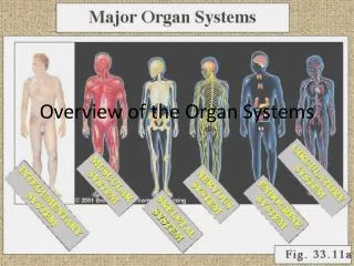
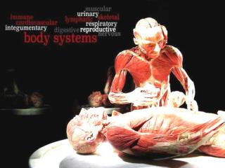
CONTENTS • HUMAN CIRCULATORY SYSTEM • HUMAN RESPIRATORY SYSTEM • HUMAN DIGESTIVE SYSTEM • HUMAN EXCRETORY SYSTEM • HUMAN REPRODUCTION SYSTEM
BRIEF INTRODUCTION • The circulatory system is an organ system that moves nutrients, gases, and wastes to and from cells, helps fight diseases and helps stabilize body temperature and pH to maintain homeostasis. This system may be seen strictly as a blood distribution network, but some consider the circulatory system as composed of the cardiovascular system, which distributes blood, and the lymphatic system, which distributes lymph. While humans, as well as other vertebrates, have a closed cardiovascular system (meaning that the blood never leaves the network of arteries, veins and capillaries), some invertebrate groups have an open cardiovascular system. The most primitive animal phyla lack circulatory systems. The lymphatic system, on the other hand, is an open system. • The main components of the human circulatory system are the heart, the blood, and the blood vessels. The circulatory system includes: the pulmonary circulation, a "loop" through the lungs where blood is oxygenated; and the systemic circulation, a "loop" through the rest of the body to provide oxygenated blood. An average adult contains five to six quarts (roughly 4.7 to 5.7 litres) of blood, which consists of plasma, red blood cells, white blood cells, and platelets. Also, the digestive system works with the circulatory system to provide the nutrients the system needs to keep the heart pumping. • Two types of fluids move through the circulatory system: blood and lymph. The blood, heart, and blood vessels form the cardiovascular system. The lymph, lymph nodes, and lymph vessels form the lymphatic system. The cardiovascular system and the lymphatic system collectively make up the circulatory system.
PULMONARY CIRCULATION • Pulmonary circulation is the portion of the cardiovascular system which transports oxygen-depleted blood away from the heart, to the lungs, and returns oxygenated blood back to the heart. • De-oxygenated blood enters the right atrium of the heart and flows into the right ventricle where it is pumped through the pulmonary arteries to the lungs. Pulmonary veins return the now oxygen-rich blood to the heart, where it enters the left atrium before flowing into the left ventricle. Also, from the left ventricle the oxygen-rich blood is pumped out via the aorta, and on to the rest of the body.
CORONARY CIRCULATION • The coronary circulatory system provides a blood supply to the heart. As it provides oxygenated blood to the heart, it is by definition a part of the systemic circulatory system.
HEART • The heart pumps oxygenated blood to the body and deoxygenated blood to the lungs. In the human heart there is one atrium and one ventricle for each circulation, and with both a systemic and a pulmonary circulation there are four chambers in total: left atrium, left ventricle, right atrium and right ventricle. The right Atrium, which is the upper chamber of the right side of the heart, receives blood from the upper body through the Superior Vena Cava, and from the lower body through the Inferior Vena Cava.
CLOSED CARDIOVASCULAR SYSTEM • The cardiovascular systems of humans are closed, meaning that the blood never leaves the network of blood vessels. In contrast, oxygen and nutrients diffuse across the blood vessel layers and enters interstitial fluid, which carries oxygen and nutrients to the target cells, and carbon dioxide and wastes in the opposite direction. The other component of the circulatory system, the lymphatic system, is not closed.
THE OPEN CIRCULATORY SYSTEM • The open circulatory system is an arrangement of internal transport present in many animals such as molluscs and arthropods, in which fluid (called hemolymph) in a cavity called the hemocoel bathes the organs directly with oxygen and nutrients and there is no distinction between blood and interstitial fluid; this combined fluid is called hemolymph or haemolymph. Muscular movements by the animal during locomotion can facilitate hemolymph movement, but diverting flow from one area to another is limited. When the heart relaxes, blood is drawn back toward the heart through open-ended pores (ostia). • Hemolymph fills all of the interior hemocoel of the body and surrounds all cells. Hemolymph is composed of water, inorganic salts (mostly Na+, Cl-, K+, Mg2+, and Ca2+), and organic compounds (mostly carbohydrates, proteins, and lipids). The primary oxygen transporter molecule is hemocyanin. • There are free-floating cells, the hemocytes, within the hemolymph. They play a role in the arthropod immune system.
BRIEF INTRODUCTION • In living organisms, a respiratory system's function is to allow gas exchange. The space between the alveoli and the capillaries, the anatomy or structure of the exchange system, and the precise physiological uses of the exchanged gases vary depending on the organism. In humans and other mammals, for example, the anatomical features of the respiratory system include airways, lungs, and the respiratory muscles. Molecules of oxygen and carbon dioxide are passively exchanged, by diffusion, between the gaseous external environment and the blood. This exchange process occurs in the alveolar region of the lungs. • Other animals, such as insects, have respiratory systems with very simple anatomical features, and in amphibians even the skin plays a vital role in gas exchange. Plants also have respiratory systems but the directionality of gas exchange can be opposite to that in animals. The respiratory system in plants also includes anatomical features such as holes on the undersides of leaves known as stomata.
ANATOMY OF RESPIRATORY SYSTEM IN VERTEBRATES • For mammals, including humans, respiration is essential. In these organisms, the respiratory system can be subdivided into an upper respiratory tract and a lower respiratory tract based on anatomical features. The upper respiratory tract includes the nasal passages, pharynx and the larynx, while the lower respiratory tract is comprised of the trachea, the primary bronchi and lungs. The respiratory system can also be divided into physiological, or functional, zones. These include the conducting zone (the region for gas transport from the outside atmosphere to just above the alveoli), the , and the respiratory zone (the alveolar region where gas exchange occurs). 1.Mammals
2.Unique aspects of comparative anatomy/physiology in mammals (i)Horses • Horses are obligate nasal breathers. That is, they are different from many other mammals in that they do not have the option of breathing through their mouths and must take in air through their nose. • The horse's respiratory system is divided into two sections, the upper respiratory tract and the lower respiratory tract. The upper respiratory tract includes the nostrils, the nasal passages, pharynx, larynx and the trachea. The lower respiratory tract is made up of the bronchi, bronchioles, and alveoli, all of which reside within the lungs of the horse. (ii)Elephants • The elephant is the only mammal known to have no pleural space. Rather, the parietal and visceral pleura are both composed of dense connective tissue and joined to each other via loose connective tissue. This lack of a pleural space, along with an unusually thick diaphragm, are thought to be evolutionary adaptations allowing the elephant to remain underwater for long periods of time while breathing through its trunk which emerges as a snorkel.
3.Birds • The respiratory system of birds differs significantly from that found in mammals, containing unique anatomical features such as air sacs. The lungs of birds also do not have the capacity to inflate as birds lack a diaphragm and a pleural cavity. Gas exchange in birds occurs between air capillaries and blood capillaries, rather than in alveoli. 4.Reptiles • The anatomical structure of the lungs is less complex in reptiles than in mammals, with reptiles lacking the extensive airway tree structure found in mammalian lungs. Gas exchange in reptiles still occurs in alveoli, however. Reptiles do not possess a diaphragm. Thus, breathing occurs via a change in the volume of the body cavity which is controlled by contraction of intercostal muscles in all reptiles except turtles. In turtles, contraction of specific pairs of flank muscles governs inspiration or expiration.
5.Amphibians • The skin is one of the important respiratory organs in amphibians. It is highly vascularized and moist, with moisture maintained via secretion of mucus from specialized cells. These properties aid rapid gas exchange. • In most fish the respiration takes place through gills. Lungfish, however, do possess one or two lungs. The labyrinth fishes have developed a special organ that allows them to take advantage of the oxygen of the air, but is not a true lung. The rare breath fish also has two lungs. 6.Fish
ANATOMY OF RESPIRATORY SYSTEM IN INVERTEBRATES • These animals lack specialized organs for gas exchange, instead taking in gases directly from the surrounding water. 1.Sponges and jellyfish 2.Flatworms and annelids • Flatworms have special muscles, called "{Lang|lat|enmmustullus}}", meaning "small muscles" in Latin. These muscles help the worms to create energy efficiently, while still completing essential activities like eating and sleeping.
3.Insects • Air enters the respiratory system of most insects through a series of external openings called spiracles. These external openings, which act as muscular valves in some insects, lead to the internal respiratory system, a densely-networked array of tubes called trachea. The tracheal system within an individual is composed of interconnecting transverse and longitudinal tracheae which maintain equivalent pressure throughout the system. These tracheae branch repeatedly, eventually forming tracheoles, which are blind-ended, water-filled compartments only one micrometer in diameter. It is at this level of the tracheoles that oxygen is delivered to the cells for respiration. • Insects were once believed to exchange gases with the environment continuously by the simple diffusion of gases into the tracheal system. More recently, however, large variation in insect ventilatory patterns have been documented and insect respiration appears to be highly variable. Some small insects do demonstrate continuous respiration and may lack muscular control of the spiracles. Others, however, utilize muscular contraction of the abdomen along with coordinated spiracle contraction and relaxation to generate cyclical gas exchange patterns. The most extreme form of these patterns is termed discontinuous gas exchange cycles (DGC).
PHYSIOLOGY OF RESPIRATORY SYSTEM IN MAMMALS 1.Ventilation • Ventilation occurs under the control of the autonomic nervous system from parts of the brain stem, the medulla oblongata and the pons. This area of the brain forms the respiration regulatory center, a series of interconnected brain cells within the lower and middle brain stem which coordinate respiratory movements. The sections are the pneumotaxic center, the apneustic center, and the dorsal and ventral respiratory groups. This section is especially sensitive during infancy, and the neurons can be destroyed if the infant is dropped and/or shaken violently. The result can be death due to "shaken baby syndrome." • Ventilation of the lungs is carried out by the muscles of respiration. 2.Control
3.Inhalation • Inhalation is initiated by the diaphragm and supported by the external intercostal muscles. Normal resting respirations are 10 to 18 breaths per minute, with a time period of 2 seconds. During vigorous inhalation (at rates exceeding 35 breaths per minute), or in approaching respiratory failure, accessory muscles of respiration are recruited for support. These consist of sternocleidomastoid, platysma, and the scalene muscles of the neck. • Under normal conditions, the diaphragm is the primary driver of inhalation. When the diaphragm contracts, the ribcage expands and the contents of the abdomen are moved downward. This results in a larger thoracic volume and negative (suction) pressure (with respect to atmospheric pressure) inside the thorax. As the pressure in the chest falls, air moves into the conducting zone. Here, the air is filtered, warmed, and humidified as it flows to the lungs. • During forced inhalation, as when taking a deep breath, the external intercostal muscles and accessory muscles aid in further expanding the thoracic cavity. 4.Exhalation • Exhalation is generally a passive process; however, active or forced exhalation is achieved by the abdominal and the internal intercostal muscles. During this process air is forced or exhaled out. • The lungs have a natural elasticity: as they recoil from the stretch of inhalation, air flows back out until the pressures in the chest and the atmosphere reach equilibrium. • During forced exhalation, as when blowing out a candle, expiratory muscles including the abdominal muscles and internal intercostal muscles, generate abdominal and thoracic pressure, which forces air out of the lungs.
5.Circulation • The right side of the heart pumps blood from the right ventricle through the pulmonary semilunar valve into the pulmonary trunk. The trunk branches into right and left pulmonary arteries to the pulmonary blood vessels. The vessels generally accompany the airways and also undergo numerous branchings. Once the gas exchange process is complete in the pulmonary capillaries, blood is returned to the left side of the heart through four pulmonary veins, two from each side. The pulmonary circulation has a very low resistance, due to the short distance within the lungs, compared to the systemic circulation, and for this reason, all the pressures within the pulmonary blood vessels are normally low as compared to the pressure of the systemic circulation loop. 6.Gas exchange • The major function of the respiratory system is gas exchange between the external environment and an organism's circulatory system. In humans and mammals, this exchange facilitates oxygenation of the blood with a concomitant removal of carbon dioxide and other gaseous metabolic wastes from the circulation. As gas exchange occurs, the acid-base balance of the body is maintained as part of homeostasis. If proper ventilation is not maintained, two opposing conditions could occur: 1) respiratory acidosis, a life threatening condition, and 2) respiratory alkalosis. • Upon inhalation, gas exchange occurs at the alveoli, the tiny sacs which are the basic functional component of the lungs. The alveolar walls are extremely thin (approx. 0.2 micrometers). These walls are composed of a single layer of epithelial cells (type I and type II epithelial cells) in close proximity to the pulmonary capillaries which are composed of a single layer of endothelial cells. The close proximity of these two cell types allows permeability to gases and, hence, gas exchange.
7.Non respiratory functions (i)Vocalization • The movement of gas through the larynx, pharynx and mouth allows humans to speak, or phonate. Vocalization, or singing, in birds occurs via the syrinx, an organ located at the base of the trachea. The vibration of air flowing across the larynx (vocal chords), in humans, and the syrinx, in birds, results in sound. Because of this, gas movement is extremely vital for communication purposes. (ii)Temperature control • Panting in dogs and other animals that are mammals need Temperature control. (iii)Coughing and sneezing • The phlegm is removed from the body by coughing, and sneezing and irritated nerves within nasal passages.
DEVELOPMENT OF RESPIRATORY SYSTEM IN ANIMALS • The respiratory system lies dormant in the human fetus during pregnancy. At birth, the respiratory system becomes fully functional upon exposure to air, although some lung development and growth continues throughout childhood. Pre-term birth can lead to infants with under-developed lungs. These lungs show incomplete development of the alveolar type II cells, cells that produce surfactant. The lungs of pre-term infants may not function well because the lack of surfactant leads to increased surface tension within the alveoli. Thus, many alveoli collapse such that no gas exchange can occur within some or most regions of an infant's lungs, a condition termed respiratory distress syndrome. Basic scientific experiments, carried out using cells from chicken lungs, support the potential for using steroids as a means of furthering development of type II alveolar cells. In fact, once a pre-mature birth is threatened, every effort is made to delay the birth, and a series of steroid shots is frequently administered to the mother during this delay in an effort to promote lung growth.
DISEASE AND RESPIRATORY SYSTEM Disorders of the respiratory system can be classified into four general areas: • Obstructive conditions (e.g., emphysema, bronchitis, asthma attacks) • Restrictive conditions (e.g., fibrosis, sarcoidosis, alveolar damage, pleural effusion) • Vascular diseases (e.g., pulmonary edema, pulmonary embolism, pulmonary hypertension) • Infectious, environmental and other "diseases" (e.g., pneumonia, tuberculosis, asbestosis, particulate pollutants): Coughing is of major importance, as it is the body's main method to remove dust, mucus, saliva, and other debris from the lungs. Inability to cough can lead to infection. Deep breathing exercises may help keep finer structures of the lungs clear from particulate matter, etc. The respiratory tract is constantly exposed to microbes due to the extensive surface area, which is why the respiratory system includes many mechanisms to defend itself and prevent pathogens from entering the body. Disorders of the respiratory system are usually treated internally by a pulmonologist or respiratory physician.
BRIEF INTRODUCTION • Digestion is the breaking down of chemicals in the body, into a form that can be absorbed. It is also the process by which the body breaks down chemicals into smaller components that can be absorbed by the blood stream. In mammals, preparation for digestion begins with the cephalic phase in which saliva is produced in the mouth and digestive enzymes are produced in the stomach. Mechanical and chemical digestion begin in the mouth where food is chewed, and mixed with saliva to break down starches. The stomach continues to break food down mechanically and chemically through the churning of the stomach and mixing with enzymes. Absorption occurs in the stomach and gastrointestinal tract, and the process finishes with defecation.
OVERVIEW In most vertebrates, digestion is a multi-stage process in the digestive system, starting from ingestion of raw materials, most often other organisms. The process of ingestion usually inolves some type of mechanical and chemical processing. Digestion is separated into four steps: • Ingestion: placing food into the mouth(entry of food in the digestive system) • Mechanical and chemical breakdown: mastication and the mixing of the resulting food bolus with water, acids, bile and enzymes in the stomach and intestine to break down complex molecules into simple structures • Absorption: of nutrients from the digestive system to the circulatory and lymphatic capillaries through osmosis, active transport, and diffusion • Egestion: Removal of undigested materials from the digestive tract through defecation Underlying the process is muscle movement throughout the system through swallowing and peristalsis. The major part of the digestive process takes place in the small intestine. The stomach takes in the food, then churns it and makes it into very small particles called "chime."
HUMAN DIGESTION PROCESS Phases of gastric secretion • Cephalic phase - This phase occurs before food enters the stomach and involves preparation of the body for eating and digestion. Sight and thought stimulate the cerebral cortex. Taste and smell stimulus is sent to the hypothalamus and medulla oblongata. After this it is routed through the vagus nerve and release of acetylcholine. Gastric secretion at this phase rises to 40% of maximum rate. Acidity in the stomach is not buffered by food at this point and thus acts to inhibit parietal (secretes acid) and G cell (secretes gastrin) activity via D cell secretion of somatostatin. • Gastric phase - This phase takes 3 to 4 hours. It is stimulated by distention of the stomach, presence of food in stomach and increase in pH. Distention activates long and myentric reflexes. This activates the release of acetylcholine which stimulates the release of more gastric juices. As protein enters the stomach, it binds to hydrogen ions, which raises the pH of the stomach to around pH 6. Inhibition of gastrin and HCl secretion is lifted. This triggers G cells to release gastrin, which in turn stimulates parietal cells to secrete HCl. HCl release is also triggered by acetylcholine and histamine. • Intestinal phase - This phase has 2 parts, the excitatory and the inhibitory. Partially-digested food fills the duodenum. This triggers intestinal gastrin to be released. Enterogastric reflex inhibits vagal nuclei, activating sympathetic fibers causing the pyloric sphincter to tighten to prevent more food from entering, and inhibits local reflexes.
Oral cavity • In humans, digestion begins in the oral cavity where food is chewed. Saliva is secreted in large amounts (1-1.5 litres/day) by three pairs of exocrine salivary glands (parotid, submandibular, and sublingual) in the oral cavity, and is mixed with the chewed food by the tongue. There are two types of saliva. One is a thin, watery secretion, and its purpose is to wet the food. The other is a thick, mucous secretion, and it acts as a lubricant and causes food particles to stick together and form a bolus. The saliva serves to clean the oral cavity and moisten the food, and contains digestive enzymes such as salivary amylase, which aids in the chemical breakdown of polysaccharides such as starch into disaccharides such as maltose. It also contains mucin, a glycoprotein which helps soften the food into a bolus. • Swallowing transports the chewed food into the esophagus, passing through the oropharynx and hypopharynx. The mechanism for swallowing is coordinated by the swallowing center in the medulla oblongata and pons. The reflex is initiated by touch receptors in the pharynx as the bolus of food is pushed to the back of the mouth.
Esophagus • The esophagus is a narrow muscular tube about 25 centimeters long which starts at pharynx at the back of the mouth, passes through the thorax and thoracic diaphragm, and ends at the cardiac orifice of the stomach. The wall of the esophagus is made up of two layers of smooth muscles, which form a continuous layer from the esophagus to the oten and contract slowly, over long periods of time. The inner layer of muscles is arranged circularly in a series of descending rings, while the outer layer is arranged longitudinally. At the top of the esophagus, is a flap of tissue called the epiglottis that closes during swallowing to prevent food from entering the trachea (windpipe). The chewed food is pushed down the esophagus to the stomach through peristaltic contraction of these muscles. It takes only about seven seconds for food to pass through the esophagus and no digestion takes place.
Stomach The stomach is a small,'C'-shaped pouch with walls made of thick, elastic muscles, which stores and helps break down food. Food enters the stomach through the cardiac orifice where it is further broken apart and thoroughly mixed with gastric acid, pepsin and other digestive enzymes to break down proteins. The acid itself does not break down food molecules, rather it provides an optimum pH for the reaction of the enzyme pepsin and kills many microorganisms that are ingested with the food. The parietal cells of the stomach also secrete a glycoprotein called intrinsic factor which enables the absorption of vitamin B-12. Other small molecules such as alcohol are absorbed in the stomach, passing through the membrane of the stomach and entering the circulatory system directly. Food in the stomach is in semi-liquid form. The transverse section of the alimentary canal reveals four distinct and well developed layers within the stomach: • Serous membrane, a thin layer of mesothelial cells that is the outermost wall of the stomach. • Muscular coat, a well-developed layer of muscles used to mix ingested food, composed of three sets running in three different alignments. The outermost layer runs parallel to the vertical axis of the stomach (from top to bottom), the middle is concentric to the axis (horizontally circling the stomach cavity) and the innermost oblique layer, which is responsible for mixing and breaking down ingested food, runs diagonal to the longitudinal axis. The inner layer is unique to the stomach, all other parts of the digestive tract have only the first two layers. • Submucosa, composed of connective tissue that links the inner muscular layer to the mucosa and contains the nerves, blood and lymph vessels. • Mucosa is the extensively folded innermost layer filled with connective tissue and covered in gastric glands that may be simple or branched tubular, and secret mucus, hydrochloric acid, pepsinogen and renin. The mucus lubricates the food and also prevents hydrochloric acid from acting on the walls of the stomach.
Small intestine • After being processed in the stomach, food is passed to the small intestine via the pyloric sphincter. The majority of digestion and absorption occurs here after the milky chyme enters the duodenum. Here it is further mixed with three different liquids: • Bile, which emulsifies fats to allow absorption, neutralizes the chyme and is used to excrete waste products such as bilin and bile acids. • Pancreatic juice made by the pancreas. • Intestinal enzymes of the alkaline mucosal membranes. The enzymes include maltase, lactase and sucrase (all three of which process only sugars), trypsin and chymotrypsin. • As the pH level changes in the small intestines and gradually becomes basic, more enzymes are activated further that chemically break down various nutrients into smaller molecules to allow absorption into the circulatory or lymphatic systems. Small, finger-like structures called vill, each of which is covered with even smaller hair-like structures called microvilli improve the absorption of nutrients by increasing the surface area of the intestine and enhancing speed at which nutrients are absorbed. Blood containing the absorbed nutrients is carried away from the small intestine via the hepatic portal vein and goes to the liver for filtering, removal of toxins, and nutrient processing. • The small intestine and remainder of the digestive tract undergoes peristalsis to transport food from the stomach to the rectum and allow food to be mixed with the digestive juices and absorbed. The circular muscles and longitudinal muscles are antagonistic muscles, with one contracting as the other relaxes. When the circular muscles contract, the lumen becomes narrower and longer and the food is squeezed and pushed forward. When the longitudinal muscles contract, the circular muscles relax and the gut dilates to become wider and shorter to allow food to enter.
Large intestine • After the food has been passed through the small intestine, the food enters the large intestine. The large intestine is roughly 1.5 meters long, with three parts: the cecum at the junction with the small intestine, the colon, and the rectum. The colon itself has four parts: the ascending colon, the transverse colon, the descending colon, and the sigmoid colon. The large intestine absorbs water from the bolus and stores feces until it can be egested. Food products that cannot go through the villi, such as cellulose (dietary fiber), are mixed with other waste products from the body and become hard and concentrated feces. The feces is stored in the rectum for a certain period and then the stored feces is egested due to the contraction and relaxation through the anus. The exit of this waste material is regulated by the anal sphincter.
Fat digestion • The presence of fat in the small intestine produces hormones which stimulate the release of lipase from the pancreas and bile from the gallbladder. The lipase (activated by acid) breaks down the fat into monoglycerides and fatty acids. The bile emulsifies the fatty acids so they may be easily absorbed. • Short- and some medium chain fatty acids are absorbed directly into the blood via intestine capillaries and travel through the portal vein just as other absorbed nutrients do. However, long chain fatty acids and some medium chain fatty acids are too large to be directly released into the tiny intestinal capillaries. Instead they are absorbed into the fatty walls of the intestine villi and reassembled again into triglycerides. The triglycerides are coated with cholesterol and protein (protein coat) into a compound called a chylomicron. • Within the villi, the chylomicron enters a lymphatic capillary called a lacteal, which merges into larger lymphatic vessels. It is transported via the lymphatic system and the thoracic duct up to a location near the heart (where the arteries and veins are larger). The thoracic duct empties the chylomicrons into the bloodstream via the left subclavian vein. At this point the chylomicrons can transport the triglycerides to where they are needed.
Digestive hormones There are at least four hormones that aid and regulate the digestive system: • Gastrin - is in the stomach and stimulates the gastric glands to secrete pepsinogen(an inactive form of the enzyme pepsin) and hydrochloric acid. Secretion of gastrin is stimulated by food arriving in stomach. The secretion is inhibited by low pH . • Secretin - is in the duodenum and signals the secretion of sodium bicarbonate in the pancreas and it stimulates the bile secretion in the liver. This hormone responds to the acidity of the chyme. • Cholecystokinin (CCK) - is in the duodenum and stimulates the release of digestive enzymes in the pancreas and stimulates the emptying of bile in the gall bladder. This hormone is secreted in response to fat in chyme. • Gastric inhibitory peptide (GIP) - is in the duodenum and decreases the stomach churning in turn slowing the emptying in the stomach. Another function is to induce insulin secretion.
Significance of pH in digestion • Digestion is a complex process which is controlled by several factors. pH plays a crucial role in a normally functioning digestive tract. In the mouth, pharynx, and esophagus, pH is typically about 6.8, very weakly acidic. Saliva controls pH in this region of the digestive tract. Salivary amylase is contained in saliva and starts the breakdown of carbohydrates into monosaccharides. Most digestive enzymes are sensitive to pH and will not function in a low-pH environment like the stomach. A pH below 7 indicates an acid, while a pH above 7 indicates a base; the concentration of the acid or base, however, does also play a role. • pH in the stomach is very acidic and inhibits the breakdown of carbohydrates while there. The strong acid content of the stomach provides two benefits, both serving to denature proteins for further digestion in the small intestines, as well as providing non-specific immunity, retarding or eliminating various pathogens. • In the small intestines, the duodenum provides critical pH balancing to activate digestive enzymes. The liver secretes bile into the duodenum to neutralise the acidic conditions from the stomach. Also the pancreatic duct empties into the duodenum, adding bicarbonate to neutralize the acidic chyme, thus creating a neutral environment. The mucosal tissue of the small intestines is alkaline, creating a pH of about 8.5, thus enabling absorption in a mild alkaline in the environment.
BRIEF INTRODUCTION • The excretory system excretes wastes. It is responsible for the elimination of the nitrogenous waste products of metabolism as well as other non-useful nitrogenous materials. Lungs are two organs localized on each side of the thorax. They are constituted by pulmonary alveoli. They are responsible for converting oxygen into carbon dioxide, but to maintain the organism's cells can take the oxygen through passive diffusion from the bloodstream and use it in its own metabolism, thus producing carbon dioxide that will be further excreted when passing through alveoli circulation. • The process by which animals rid themselves of waste products and of the nitrogenous by-products of metabolism. Through excretion organisms control osmotic pressure—the balance between inorganic ions and water—and maintain acid-base balance. The process thus promotes homeostasis, the constancy of the organism's internal environment. • Every organism, from the smallest protista to the largest mammal, must rid itself of the potentially harmful by-products of its own vital activities. This process in living things is called elimination, which may be considered to encompass all of the various mechanisms and processes by which life forms dispose of or throw off waste products, toxic substances, and dead portions of the organism. The nature of the process and of the specialized structures developed for waste complexity of the organism. • Four terms are commonly associated with waste-disposal processes and are often used interchangeably, though not always correctly: excretion, secretion, egestion, and elimination. • Excretion is a general term referring to the separation and throwing off of waste materials or toxic substances from the cells and tissues of a plant or animal.
The separation, elaboration, and elimination of certain products arising from cellular functions in multicellular organisms is called secretion. Though these substances may be a waste product of the cell producing them, they are frequently useful to other cells of the organism. Examples of secretions are the digestive enzymes produced by intestinal and pancreatic tissue cells of vertebrate animals, the hormones synthesized by specialized glandular cells of plants and animals, and sweat secreted by glandular cells in the skins of some mammals. Secretion implies that the chemical compounds being secreted were synthesized by specialized cells and that they are of functional value to the organism. The disposal of common waste products should not, therefore, be considered to be of a secretory nature. • Egestion is the act of excreting unusable or undigested material from a cell, as in the case of single-celled organisms, or from the digestive tract of multicellular animals. • As defined above, elimination broadly defines the mechanisms of waste disposal by living systems at all levels of complexity. The term may be used interchangeably with excretion. The waste then travels to anus and is released. SKIN • The skin is another part of the excretory system, containing sweat that help regulate the concentration in one’s body while also keeping him or her cool. The salt helps evaporate the water, cooling off the skin. Sweat is excreted through sweat glands. There are two types of sweat glands: eccrine sweat glands and apocrine sweat glands. The basic purpose of skin is to provide a waterproof, protective, covering for the body's complex internal environment. The skin also plays a key role in helping to maintain the circulatory and nervous system.
ECCRINE SWEAT GLANDS • The eccrine sweat glands secrete mostly water and salt and are used by the body for temperature control. These glands are located all around the body but are most profuse around the soles of the feet, palms of the hands, and the forehead. LIVER • The liver is an accessory of the digestive system. It weighs more than 1.5 kg. It is the body's largest and most complicated organ and is shaped like a pyramid. It also helps in excreting wastes from the body. • The liver absorbs drugs and other poisonous substances in the blood. It takes part in several hundred chemical reactions that control the composition of the blood and supply the needs of other organs. It changes the chemical structure of these substances are then excreted in the bile. The bile is secretion of the liver. It makes digestion of fats easier and also carries away waste production.
KIDNEYS • The most important organs of the excretory system are the kidneys. The kidneys are placed on either side of the spinal column near the lower back. The kidneys are bean-shaped and they have an important job. They are responsible for removing wastes from the blood and they also keep your blood pressure in check and help with the making of red blood cells. The kidneys filter the blood and remove any wastes. The Kidney does this via its three layers which are the Cortex, the medulla and the pelvis. In the Cortex and Medulla there are Nephrons. These Nephrons comprise of a Glomerulus (bundle of capillaries), a Bowman's Capsule, a Proximal Convoluted Tubuale, the descending and ascending Loop of Henle, the Distal Convoluted Tubual and Collecting Ducts. The collecting ducts come together in the Pelvis. When your body gets ready to pass waste products, it goes through the kidneys and mixes with water and urine. Then, the waste travels into the bladder through tubes. These tubes are called Ureters. Now, the bladder holds all of that urine until it feels so full that you need to get rid of it. That's called urination. When this happens, a tube called the Urethra takes the urine to the outside of the body. It also produces Bile.
BILE • Bile is a substance secreted by the abdomen and used for breaking wastes, with the help of estrogen. It is composed of water, cholesterol, lecithin, bicarbonate ions, bile salts, and pigments. Bile is a strong basic substance, classified as alkaline. It is released through bile ducts in the liver. Bile is a digestive chemical that is produce in the liver, stored in the gall bladder, and secreted in the small intestine. The kidney's main roles are to control the blood PH, filter all kind of wastes and remove urea from the blood and produced in to urine. Bile is capable of breaking down fats.
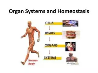
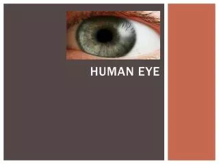
Female Reproductive System Male Reproductive System HUMAN REPRODUCTIVE SYSTEM
BRIEF INTRODUCTION • The reproductive system is a system of organs within an organism which work together for the purpose of reproduction. Many non-living substances such as fluids, hormones, and pheromones are also important accessories to the reproductive system. Unlike most organ systems, the sexes of differentiated species often have significant differences. These differences allow for a combination of genetic material between two individuals, which allows for the possibility of greater genetic fitness of the offspring. • The major organs of the human reproductive system include the external genitalia (penis and vulva) as well as a number of internal organs including the gamete producing gonads (testicles and ovaries). Diseases of the human reproductive system are very common and widespread, particularly communicable sexually transmitted diseases. • Most other vertebrate animals have generally similar reproductive systems consisting of gonads, ducts, and openings. However, there is a great diversity of physical adaptations as well as reproductive strategies in every group of vertebrates.
HUMAN REPRODUCTIVE SYSTEM • Human reproduction takes place as internal fertilization by sexual intercourse. During this process, the erect penis of the male is inserted into the female's vagina until the male ejaculates semen, which contains sperm, into the female's vagina. The sperm then travels through the vagina and cervix into the uterus or fallopian tubes for fertilization of the ovum. Upon successful fertilization and implantation, gestation of the foetus then occurs within the female's uterus for approximately nine months, this process is known as pregnancy in humans. Gestation ends with birth, the process of birth is known as labor. Labor consists of the muscles of the uterus contracting, the cervix dilating, and the baby passing out the vagina. Human's babies and children are nearly helpless and require high levels of parental care for many years. One important type of parental care is the use of the mammary glands in the female breasts to nurse the baby. • Humans have a high level of sexual differentiation. In addition to differences in nearly every reproductive organ, numerous differences typically occur in secondary sexual characteristics and in sexual and parental behaviours.
Male reproductive system • The human male reproductive system is a series of organs located outside of the body and around the pelvic region of a male that contribute towards the reproductive process. The primary direct function of the male reproductive system is to provide the male gamete or spermatozoa for fertilization of the ovum. • The major reproductive organs of the male can be grouped into three categories. The first category is sperm production and storage. Production takes place in the testes which are housed in the temperature regulating scrotum, immature sperm then travel to the epididymis for development and storage. The second category are the ejaculatory fluid producing glands which include the seminal vesicles, prostate, and the vas deferens. The final category are those used for copulation, and deposition of the spermatozoa (sperm) within the female, these include the penis, urethra, vas deferens, and Cowper's gland. • Major secondary sexual characteristics include: larger, more muscular stature, deepened voice, facial and body hair and broad shoulders. An important sexual hormone of males is androgen, and particularly testosterone.