Download
1 / 36
370 likes | 654 Views
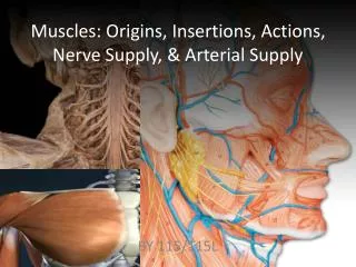
Nerve Supply of the UL. Dr: Yasser S eddeg. BRACHIAL PLEXUS. FORMATION of the Brachial Plexus. ROOTS- C5-C8 AND TI( VENTRAL RAMI) TRUNKS- UPPER , MIDDLE, LOWER DIVISONS- ANTERIOR/POSTERIOR CORDS- MEDIAL/LATERAL/POSTERIOR. ROOTS FORMATION OF TRUNKS C 5 C 6 C 7 C 8 T 1.

E N D
Nerve Supply of the UL Dr: Yasser Seddeg
FORMATION of the Brachial Plexus • ROOTS- C5-C8 AND TI( VENTRAL RAMI) • TRUNKS-UPPER , MIDDLE, LOWER • DIVISONS-ANTERIOR/POSTERIOR • CORDS-MEDIAL/LATERAL/POSTERIOR
ROOTS FORMATION OF TRUNKS • C5 • C6 • C7 • C8 • T1 UPPER MIDDLE LOWER
DIVISIONS AND CORDS OF BRACHIAL PLEXUS • TRUNKS DIVISIONS CORDS • UPPER TRUNK • MIDDLE TRUNK • LOWER TRUNK LATERAL CORD-ANTERIOR DIVISIONS OF UPPER AND MIDDLE TRUNK POSTERIOR CORD-POSTERIORDIVISIONS OF ALL TRUNKS MEDIAL CORD-ANTERIOR DIVISION OF LOWER TRUNK CORDS ARE NAMED ACCORDING TO THEIR RELATIVE POSITIONS TO THE AXILLARY ARTERY anterior posterior anterior posterior anterior posterior
NERVES ARISING FROM THE CORDS • MEDIAL CORD • MEDIAL CUTANEOUS NERVE OF ARM- CUTANEOUS SUPPLY TO SKIN • MEDIAL CUTANEOUS NERVE OF FOREARM-CUTANEOUS SUPPLY TO THE SKIN • MEDIAL ROOT OF MEDIAL NERVE- JOINS WITH LATERAL ROOT TO FORM MEDIAN NERVE • ULNAR NERVE • MEDIAL PECTORAL NERVE
NERVES FROM THE LATERAL CORD • LATERAL ROOT OF MEDIAN NERVE • MUSCULOCUTANEOUS NERVE • LATERAL PECTORAL NERVE
NERVES FROM THE POSTERIOR CORD • 1. UPPER SUBSCAPULAR NERVE • 2.LOWER SUBSCAPULAR NERVE • 3.THORACODORSAL NERVE • 4. RADIAL NERVE • 5. AXILLARY NERVE
Muscles of Scapula Rotator Cuff • If ORIGIN on scapula = Move Arm • Subscapularis • Supraspinatus • Infraspinatus • Teres Minor • Teres Major • Latissimus Dorsi (partial O on scap) • Coracobrachialis • If INSERTION on scapula = Move scapula • Rhomboids • Trapezius • Pectoralis Minor • Serratus Ventralis • Levator Scapulae
Innervation of Scapula Muscles • Origin on Scapula: • Latissimusdorsi = Thoracodorsal nerve • Subscapularis, Teres Major = Subscapular nerves • Supraspinatus, Infraspinatus = Suprascapular nerves • Teres Minor = Axillary nerve • Insertion on Scapula • Levator Scapular, Rhomboids = Dorsal Scapular nerve • Pectoralis Minor = Pectoral n. • Serratus anterior = Long Thoracic n. • Trapezius = Accessory n.
Muscles of Arm: Cross elbow, Move forearm • 2 Compartments • Anterior: Flexors of forearm • Posterior: Extensors of forearm • Anterior Compartment • Biceps brachii = MC nerve • Brachialis = MC nerve • Brachioradialis = Radial nerve • Coracobrachialis = MC nerve • O = coracoid process of scapula • I = medial side humeral shaft • A = flex, adduct arm • Posterior Compartment • Triceps brachii = Radial nerve • Anconeus = Radial nerve MC = musculocutaneous nerve
Muscles of forearm: Cross wrist + finger joints, moves hand • Cross Wrist = flex, extend, abduct, adduct hand • Cross Fingers = flex, extend fingers • Most muscles fleshy proximally, long tendons distally • Flexor + Extensor Retinacula • wristbands keep tendons from bowing • thick, deep fascia • Anterior Flexor Compartment (Superficial + Deep layers) • Most flexors have common tendon on medial epicondyle • Contains 2 pronators • Innervated by *Median, Ulna nerves • Posterior Extensor Compartment (Superficial + Deep layers) • Innervated by Radial nerve (or branches of)
Superficial Muscles Flexor digitorum superficialis Median Flexor carpi radialis Median Pronator teres Median Palmaris longus Median Flexor carpi ulnaris Ulnar Deep Muscles Pronator quadratus Median Flexor pollicis longus Median Flexor digitorum profundusUlnar (med 1/2) Median (lat 1/2) Innervation of Anterior Compartment-Forearm Muscles Muscle Nerve
Innervation of Posterior Compartment-Forearm Muscles Muscle Nerve • Superficial • Extensor carpi radialis longus Radial • Extensor digitorum Radial • Extensor carpi ulnaris Radial • Deep • Supinator Radial • Abductor pollicis longus Radial • Extensor pollicis longus + brevis Radial • Extensor indicus Radial
Intrinsic Muscles of Hand Muscle Nerve • Pinky (little finger) • All digiti minimi Ulnar (Flexor, Abductor, Opponens) • Thumb • Abductor pollicis brevis Median • Flexor pollicis brevis Median • Opponens pollicis Median • Adductor pollicis Ulnar • Other Intrinsic Muscles • Palmar + Dorsal Interossei Ulnar • Lumbricals Median, Ulnar
pg 297 Intrinsic Muscles of Hand Palmar Interossei Dorsal Interossei Lumbricals 3rd ABduction ADDuction 2nd 4th 1st
Innervation by Posterior Cord • Radial Nerve (largest branch) • Course: Through arm, around humerus, around lateral epicondyle, then divides • Innervates: all posterior muscles of arm and forearm • Triceps brachii, anconeus, supinator, brachioradialis • Divides in forearm: • Superficial = skin of arm and dorsolateral surface of hand • Deep = extensor muscles of forearm (eg E. carpi radialis L + B) • Damage to Radial Nerve = wristdrop • Inability to extend the hand, st inability to fully extend forearm
Innervation by Posterior Cord (continued) • Axillary Nerve (runs w/ caudal humeral circumflex a.) • Innervates: • Deltoid and Teres minor (motor inn) • Capsule of shoulder, skin of shoulder (sensory inn) • Subscapular Nerve {branches of C5 + C6 rami} • Innervates: Subscapularis, Teres major • Thoracodorsal Nerve (runs w/thoracodorsal a+v) • Innervates: Latissimus dorsi
Innervation by Lateral Cord • Musculocutaneous • Course: branches to arm, distal to elbow becomes cutaneous for lateral forearm skin • Innervates • Biceps brachii, brachialis, coracobrachialis (motor inn) • Skin distal to elbow (sensory) • Suprascapular • Innervates: Supraspinatus, Infraspinatus
Innervation by both Lateral and Medial Cords • Median • Course: middle of brachial plexus, does not branch in arm, distal to elbow provides many branches to most forearm flexors, passes through carpal tunnel to hand to lateral palmar intrinsics • Innervates: most muscles of anterior forearm (motor inn) • (eg) most flexors, some intrinsics (thumb) • Innervates: skin of lateral 2/3 hand on palm side, dorsum of fingers 2+3 (sensory inn) • Nerve Damage = “Ape” Hand • Inability to Oppose Thumb
Innervation by Medial Cord • Ulnar • Course: runs along medial side of arm, behind medial epicondyle, superficial to carpal tunnel into hand, branches to supply intrinsics and skin • Innervates: • FCU and part of FDP, most intrinsics (motor inn) • Skin of medial 2/3 of hand A+P (sensory inn) • Nerve Damage: Clawhand • Inability to extend fingers at interphalangeal joints, results in permanent flexion = claw
Stretch, tear, compression or avulsion of the nerves usually after forceful lateral deviation of the head from the shoulders during delivery.).
Clinical Manifestations: Erb’spalsy caused by the disruption of the upper brachial plexus. Posture of adduction and inward rotation at the shoulder with extension and pronation at the elbow and flexion of the fingers = WAITER’S TIP
serratus anterior Paralysis • winged scapula: • Due to injury of the nerve to serratus anterior