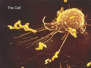
The Cell
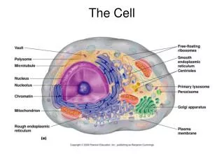
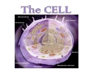
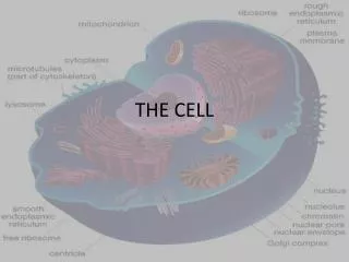
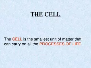
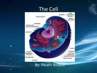
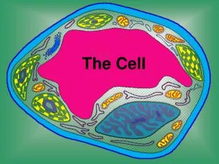
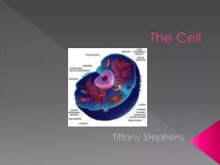
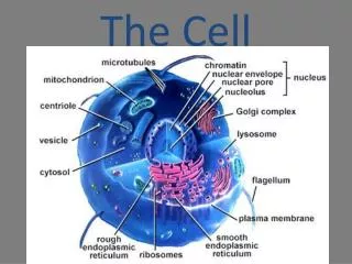
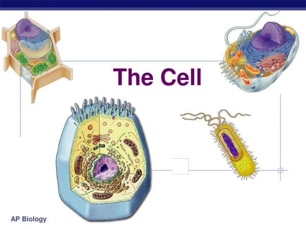
The Cell. Introduction to Cells . The basic structural and functional unit of all living things Major cellular regions The plasma membrane The cytoplasm The nucleus. *. The Plasma membrane * Structure Double layer (bilayer) of lipid molecules

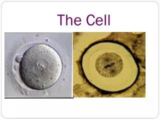
The Cell
E N D
Presentation Transcript
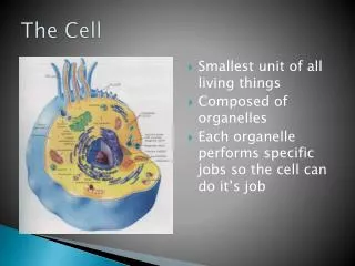
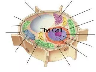
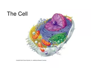
Introduction to Cells • The basic structural and functional unit of all living things • Major cellular regions • The plasma membrane • The cytoplasm • The nucleus
* • The Plasma membrane* • Structure • Double layer (bilayer) of lipid molecules • Phospholipid polar heads (hydrophilic) outward • Fatty acid chains (hydrophobic) are tail to tail • Protein molecules are dispersed within Structure of a phospholipid:
The membrane structure is actually fluid, with proteins moving around in it Plasma membrane extracellular lipid bilayer intracellular
Functions of the plasma membrane • Separates intracellular fluid from extracellular fluid • Acts as a barrier • Some membrane proteins act as receptors • Determines which substances enter and leave cell • Diffusion • Specific transport mechanisms • Bulk (vesicular) transport • Exocytosis • Endocytosis
Phagocytosis (a type of endocytosis) Receptor-mediated endocytosis Exocytosis
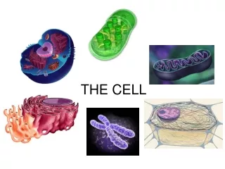
The Cytoplasm • The Cytosol: jelly-like fluid matrix • Organelles (about nine types) • Ribosomes: sites of protein synthesis • Endoplasmic reticulum (rough and smooth): products synthesized (protein, lipid, steroid); store calcium • Golgi apparatus: packages and modifies proteins • Mitochondria: synthesizes ATP (energy source) • Lysosomes: intracellular digestion (“disintegrators”) • Peroxisomes: detoxify substances • Cytoskeleton: supports cellular structures • Centrosomes and centrioles: organize microtubule network • Inclusions: not permanent (eg. food storage units and pigments)
The “Genetic Code” Replication: DNA making a copy of itself Transcription: making of RNA from code of DNA Translation: making of protein coded by tRNA via mRNA via DNA (3 bases make one protein) DNA “bases” – T, A, C, G (thymine, adenine, cytosine, guanine) RNA “bases” – U, A, C, G (uracil instead of thymine) mRNA – messenger RNA tRNA – transfer RNA rRNA - ribosomal RNA . For animation of translation: http://biology.kenyon.edu/slonc/bio3/ribo/ribo1.html Elongation of the polypeptide chain begins by the appropriate aminoacyl-tRNA binding to the codon in the A site of the ribosome.
Role of golgi apparatus in packaging products of rough ER for use in the cell and for secretion
Lysosomes Peroxisomes: like small lysosomes
The cytoskeleton: 3 types of rods (a) microtubules (b) microfilaments (c) intermediate filaments Microtubules appear as green network surrounding the cells’ blue nucleus
Centrosomes and centrioles
The nucleus • Control center of the cell • Surrounded by a nuclear envelope • Nucleolus associated with ribosome production • Chromatin - extended & condensed • DNA and histones (packaging material) • Four types of nucleotides: A, T, G, C • Nucleosomes: 8 histones wrapped in DNA • Chromosomes
NUCLEUS Control center of the cell Surrounded by a nuclear envelope Nucleolus associated with ribosome production Chromatin - extended & condensed
CHROMATIN: extended and condensed • DNA and histones (packaging material) • Four types of nucleotides: A, T, G, C • Nucleosomes: 8 histones wrapped in DNA • Chromosomes
The cell life cycle • Interphase • Variable time length • Divided into G1, S (DNA replication) and G2 subphases • Mitosis: division into two daughter cells • Interphase • Early prophase • Late prophase • Metaphase • Anaphase • Telophase and cytokinesis
Mitosis (the replicated DNA of the original cell is parceled out into 2 new cells) When chromosomes are ordered clinically, they are usually in metaphase
Developmental aspects • Human life begins as a single cell • From it, all the cells of the body will arise • All cells have the same genes yet specialization indicates differential gene activation • Cell differentiation: the development of specific and distinctive features • Aging • Cellular • Organismal
Cancer“a malignant, invasive cellular tumor that has the capacity of spreading throughout the body” • Neoplasm – “new growth” AKA tumor • Cells fail to honor normal controls of cell division • Abnormal mass of proliferating cells • Classified as • Benign – local growth • Malignant - cancer (Latin for “crab”) • Invades neighboring tissue • Can metastasize = spread • Many gene mutations may be necessary for normal cells to transform
Additional terms • Dysplasia – change in cell size, shape or arrangement; can be due to irritation; can be a precursor to cancer • Hyperplasia – increase in the number of cells • Hypertrophy – growth due to an increase in the size of the cells • Apoptosis – programmed cell death • Necrosis – death of cells or tissues because of disease or injury
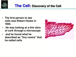
Compound Microscope All optical microscopes share the same basic components: The ocular or eyepiece (#1): to bring the image into focus for the eye. The eyepiece is inserted into the top end of the body tube. Eyepieces are interchangeable and many different eyepieces can be inserted with different degrees of magnification. Typical magnification values for eyepieces include 5x, 10x and 2x. Some eyepieces have a pointer. The objectives (#3): a cylinder containing one or more lenses, typically made of glass, to collect light from the sample. At the lower end of the microscope tube one or more objective lenses are screwed into a circular revolving nosepiece (#2) which may be rotated to select the required objective lens. Typical magnification values of objective lenses are 4x, 5x, 10x, 20x, 40x, 50x and 100x. The stage (#6): a platform below the objective which supports the specimen being viewed. In the center of the stage is a hole through which light passes to illuminate the specimen. The stage usually has arms to hold slides (rectangular glass plates with typical dimensions of 25 mm by 75 mm, on which the specimen is mounted), the mechanical stage (#9) with a control knob (#11.) The illumination source: below the stage, light is provided and controlled in a variety of ways. At its simplest, daylight is directed via a mirror. Most microscopes, however, have their own controllable light source (#7) that is focused through an optical device which concentrates it called a condenser (#8), with diaphragms (#13) controlling the amount of light let through and filters available to manage the quality and intensity of the light. The whole of the optical assembly is attached to a rigid arm (#10) which in turn is attached to a base (#12) to provide the necessary rigidity. Mounted on the arm are controls for focusing, typically a large wheel to adjust coarse focus (#4), together with a smaller one to control fine focus (#5). 10 13 11 12 http://en.wikipedia.org/wiki/Optical_Microscope