Download

1 / 59
590 likes | 849 Views
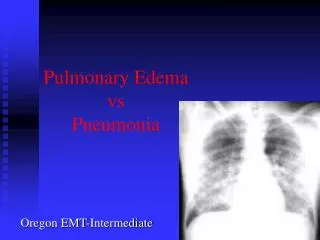
Pulmonary Edema vs Pneumonia. Paramedic Program Sp2008. Acute Pulmonary Edema, Hypotension, Shock. Clinical signs: shock, hypotension, congestive heart failure, acute pulmonary edema Most likely problem?. Acute Pulmonary Edema. Volume problem. Pump problem. Rate problem.

E N D
Pulmonary Edema vs Pneumonia Paramedic Program Sp2008
Acute Pulmonary Edema, Hypotension, Shock Clinical signs: shock, hypotension, congestive heart failure, acute pulmonary edema Most likely problem? Acute Pulmonary Edema Volume problem Pump problem Rate problem • First-line Actions • Oxygen • Nitroglycerine SL • Furosemide 0.5 to 1mg/kg • Morphine IV 2 to10 mg • Administer • Fluid Bradycardia? See algorithm Tachycardia? See algorithm Blood pressure
Let’s Review: • Cardiac Output • 5000-6000 ml/min. • HR or SV = CO • Sympathetic effects: • HR and SV • Parasympathetic: • Slows HR • Little effect on SV
Review: • SV = pressure in ventricle • Frank Starling effect • Peripheral vascular constriction increases venous return • = Increased RV output. • Vasodilation of arteries decreases PVR and diastolic pressure • = Decreased CO.
Vital Signs • Normal B/P is 120/70 mmHg • Increases with age • General: • Systolic – 100 + age up to 140 • At age 50: usually 140 mmHg • Increases 1 mmHg/yr after 50.
CHF Causes AMI Left ventricular enlargement Normal heart muscle
Dispatched as: Man down Chest pain Heart attack SOB Fainted Dizzy Passed out Choking Stroke DFO DRT Abnormal Cardiac Function
Brief History Onset Provoking factors Quality Radiation Severity Time BP changes Initial Assessment:
Initial Assessment • Meds • Cardiac rhythm • Abnormal breathing • Edema • Rales • Changes in skin color and moisture
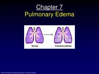
Right Heart Failure Causes COPD Left heart failure Progression Right ventricle cannot eject all of the blood Fluid/pressure backs up Right atrium Venous system Pedal edema, JVD Left Heart Failure Causes High afterload Progression Left ventricle cannot eject all of the blood Fluid/pressure backs up Left atrium Lung tissue Alveoli Pulmonary edema Right and Left Heart Failure
Acute Left Ventricular Failure • Acute LVF from heart disease: • #1 cause of heart failure. • Assume the worst, hope for best • Pt. with CAD w/ hx of MI(new or old) • May develop LVF. • Frequently LVF is only manifestation of AMI.
LVF • Common causes • Systemic HTN • Afterload • Coronary artery disease • Arteriosclerosis/atherosclerosis • Ischemia • Local/temporary occlusion
LVF • Common Causes • Infarction • Permanent, necrosis • Significant Sized Infarct • Decrease effective wall motion • Decreased stroke volume • Cardiomyopathy • Alcoholism one of main causes
LVF • Other Causes • Volume overload • Bag of Potato Chips • Severe anemia • Hypoxemia
LVF and Pulmonary Edema • Incidence of CHF doubles per decade of life • > 3 million in US; > 400,000 new diagnoses/yr • 5 yr mortality rate /p dx; • 60% in men • 43% in women
Basically this happens • Forward or backward ventricular flow. • Forward – (LVF) – reduced flow into aorta and systemic circulation • Backward – elevated systemic venous pressure
NY Heart Association’s classification of CHF • Class I • Not limited by symptoms • Class II • Fatigue, dyspnea, other sx with ordinary physical activity • Class III • Marked limitation with normal activity • Class IV • Symptoms at rest or with any activity
CHF • Acute CHF • Rapid • Chronic CHF • Slow • Midnight shoppers
Pulmonary edema also results from: • CVA • Pulmonary embolism • Infection - Sepsis • Allergy • Inhalation of fumes • Narcotic abuse • Especially Inhaled (Heroin) • Altitude sickness.
Acute Findings • History • Recent change in sleep patterns • More frequent trips to the bathroom • Need to sleep on more pillows at night • Recent move to the recliner at nights • New episodes of PND • Paroxysmal Nocturnal Dyspnea • Sudden awakening with acute shortness of breath • Relieved after standing or sitting upright for a period of time (Midnight Walmart shoppers)
Acute Findings • History • Is more nitroglycerin needed to stop the episodes of chest pain? • Have nitroglycerin or oxygen doses increased incrementally in the last few days?
Acute Findings – Critical Patient • General impression/initial assessment • Labored respirations • Audible sounds • Tripod position • Frothy sputum • Retraction of chest muscles
Acute Findings – Critical Patient • General impression/initial assessment • Lung sounds • Wheezing, crackles • Middle-to-upper lung fields • Diaphoresis, change in skin color • Severe anxiety or restlessness • Tachycardia or bradycardia • Severe hypertension may be present
Pulmonary Edema – S/S • Tachypnea • Orthopnea • Paroxysmal Nocturnal Dyspnea • Elevation of pulmonary venous & cap pressures • Wakening from sleep
Noisy Labored Breathing Fine crackles/Rales Wheezes Reflex airway spasm “Cardiac asthma” Coarse crackles/Rhonchi (larger airways) Coughing Blood Tinged Sputum Pink Frothy Pulmonary Edema – more S/S
So, What to do? • Decide – Sick/NotSick? • Vitals • Look • Skin – wet/dry, color, temp • JVD • Peripheral edema • Subtle signs
Look • Listen • Breath sounds • Pulse x 6 • Skin
Treatment of RVF & LVF • CHF a circumstance not a Dx • Treatment objectives • Decrease myocardial: • Workload • Oxygen demand • Reduce fluid retention
Treatment • Decrease Workload • No Physical activity • Sitting upright • Oxygen • Pt may tolerate BVM • CPAP – studies are promising • Decreases preload and afterload in CHF • Improves lung compliance • BiPAP • CPAP but also delivers higher pressure during inspiration
Treatment • OMI • Oxygen, Monitor, IV • MONA - if appropriate • Morphine, Oxygen, Nitro, ASA (Not in that order) • Don’t let patient walk! • Position of comfort • Reassure • Positive Pressure Ventilations if necessary
Treatment • Vasodilatory Therapy (Nitrates) • AMI reperfusion • Container expansion reduces preload • Morphine • Reduce Fluid Retention • Diuretics • Lasix • Bumex
Pneumonia Herpes Zoster Pleurisy COPD Rib fracture Asthma Angina MI Pneumothorax Pancreatitis Hepatitis Salicylate OD Bronchitis Hyperventilation Lung carcinoma Sepsis TB Muscle pain Costochondritis Pericarditis CHF Percardial tamponade Differential Diagnosis
PneumoniaThe statistics • Community acquired pneumonia • 4.5 million cases annually in US • Winter months/Colder climates • More men than women • 20% require hospitalization • 6th leading cause of death • Most common infectious cause of death
Viral • Upper and lower respiratory infections • Untreated, mortality > 30 % • 37.7% in elder > 80 y/o • Sudden onset of S/S & rapid progression suggest bacterial pneumonia
Productive cough Sputum may be Green Rust-colored Current jelly Foul smelling Rigor or shaking chills Headache Malaise N/V/D Exertional dypsnea Pleuritic chest pain, friction rub Abdominal pain S/S
Fever Tachypnea Tachycardia Cyanosis Wheezes, coarse & fine crackles Anorexia & weight loss Dullness to percussion Altered mentation S/S, cont.
nosocomial pneumonia aspiration or inhalation; ~ 45% of healthy people aspirate during sleep; even higher in severely ill patients; often bilateral typical pneumonia generally resides in the nasopharynx carried asymptomatically in approximately 50% of healthy individuals
Pneumocystis carinii pneumonia Bacterial pneumonia Bacterial pneumonia Viral pneumonia
Host Factors • DKA • Alcoholism • Sickle Cell • HIV
CHF/Pulmonary Edema Wheezes, fine & course crackles Cardiac history Productive cough ↑ dyspnea suddenly JVD Cyanosis Finger clubbing Prolonged expiratory phase Tachypnea, tachycardia Accessory muscle use Paroxysmal nocturnal dyspnea Pneumonia Wheezes, Course & fine crackles Febrile, chills Productive cough Hx URI, OM, Conjunctivitis Tachypnea, tachycardia Cyanosis H/A Malaise Abdominal distention N/V/D So – how do we tell the difference?????
Pulmonary Edema OMI MONA if approp. Position of comfort Nitroglycerin 0.4 mg SL per protocol Morphine 2-10 mg Lasix per protocol (commonly 40 mg) CPAP if available Pneumonia OMI Limit IV fluids if hx of cardiac disease CPAP if available Treatment summary
Medications for Pulmonary edema • Nitroglycerine • Morphine • Lasix
Nitroglycerin • Drug Class: Nitrate vasodilator Relieves myocardial workload • Dilates the arterial and venous systems • Reduces preload to the already overworked ventricles • Reduces blood pressure to reduce afterload • Allows pressure and fluid to move into the venous system • Sublingual doses start at 0.4mg
Morphine Sulfate • Drug Class: Narcotic Analgesic Relieves myocardial workload as well • Dilates the venous and arterial systems • Reduces preload and afterload • May cause hypotension
Morphine Sulfate: Other Actions • Mechanism of action • Binds to opiate receptors throughout the CNS • Slows respiratory rate at the medulla • Stimulates the nausea center in the brain
Morphine Sulfate • Administration • 2-4mg over 1-2 minutes, every 5 minutes (usual max dose 10 mg)