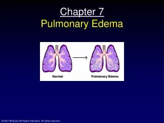
Download
1 / 30
310 likes | 738 Views
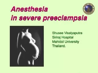
Case presentation on Pulmonary Edema Complicating Severe Preeclampsia. Presented by Dr. Nicole Hodge Faculty Advisor Dr. Norman Bolden. 6 June 06. www.anaesthesia.co.in anaesthesia.co.in@gmail.com. Overview. Severe preeclampsia – pathophysiology, Dx, maternal/fetal issues, Treatment

E N D
Case presentation on Pulmonary Edema Complicating Severe Preeclampsia Presented by Dr. Nicole Hodge Faculty Advisor Dr. Norman Bolden 6 June 06 www.anaesthesia.co.inanaesthesia.co.in@gmail.com
Overview • Severe preeclampsia – pathophysiology, Dx, maternal/fetal issues, Treatment • Standards of care and goals of anesthetic management • Case - Ante partum flash Pulmonary edema • Discussion
PREECLAMPSIA A syndrome characterized by the new onset of hypertension and proteinuria after 20 weeks gestation. Additional signs and symptoms that can occur include edema, visual disturbances, headache, epigastric pain, thrombocytopenia, and abnormal liver function. These clinical manifestations are the results of mild to severe microangiopathy of target organs such as brain, liver, kidney, and placenta.
PATHOPHYSIOLOGY OF PREECLAMPSIA A state of endothelial dysfunction secondary to excessive amounts of circulating factors released from the diseased placenta. These factors effect the establishment of a suitable vascular network of the placenta needed to supply oxygen and nutrients to the fetus. • Molecular/Cellular level • Abnormal expression of VEGF and sFlt-1 (Vascular endothelial growth factor –proangiogenic and soluble fms-like tyrosine kinase 1- anti-angiogenic factors respectively) appear to play a central role. • Increased expression of cytokines, angiotensin, catecholamines, and pro-coagulant factors. • Anatomic level • Increased vascular tone • Increased vascular permeability • Coagulopathy • Ischemia of target organs (brain, liver, kidney, placenta)
Multisystemic Disease CNS – Cortical blindness, cerebral edema, cerebral hemorrhage, and seizures Cardiovascular – Hypovolemia, increased SVR, LVH, increase sensitivity to catecholamines, sympathomimetics, and oxytocin Respiratory – pulmonary edema, V/Q mistmatch, airway edema Renal – Decreased renal blood flow, increased GFR, proteinuria, increased BUN and creatinine Hepatic – subscabular hemorrhage, abnormal LFTs, decreased plasma cholinesterase levels Hematologic – Prolonged bleeding time, platelet dysfunction, thrombocytopenia, DIC Placenta – Uteroplacental insufficiency, placental abruption, chronic fetal hypoxia, IUGR, premature labor, premature birth.
Diagnosis of Severe Preeclampsia If one or more of the following criteria are present: • Systolic blood pressure > 160 mmHg • Diastolic blood pressure greater than 110 mmHg • Proteinuria greater than 5 g/24 hrs • Evidence of end organ damage • Oliguria (<500ml/24hr) • Cerebral or visual disturbances • Pulmonary edema or cyanosis • Epigastric pain or right upper-quadrant pain • Impaired liver function • Fetal growth restriction • Thrombocytopenia *ACOG Compendium of Selected Publications
Goals The goal of the anesthesiologist • Control CNS irritability • Magnesium sulfate – anti-convulsant; reduces irritability of the neuromuscular jxn. • Restore intravasuclar fluid volume • Strictly monitor urine output • CVP monitor with goal 4-6 cm H20 • Normalize blood pressure • Magnesium sulfate – direct vasodilating action on smooth muscles of arterioles and uterus. • Labetolol, Hydralazine, nifedipine, SNP (in extreme circumstances due to fetus susceptability to cyanide toxicity) • Correct coagulation abnormalities • Platelets, FFP, Cryoprecipitate
Effects of Increasing Plasma Magnesium Levels • MgSO4 in excess of therapeutic range • Skeletal muscle weakness • Respiratory depression • Cardiac arrest (Ca++ can counter-act this) • MgSO4 potentiates NMB and sedative effects of opiods Observed Condition mEq/L Normal Plasma level 1.5-2.0 Therapeutic Range 4.0-8.0 ECG Changes (Prolonged P-Q, widened QRS) 5.0-10 Loss of deep tendon reflexes 10 SA and AV node block 15 Respiratory Paralysis 15 Cardiac Arrest 25
Anatomical Effects Functional Effects Functional effects Increased respiratory drive Airway edema friability Minimal change in TLC Increased Minute ventilation Reduced FRC Widened AP and Transverse diameter Increased cardiac output Elevated Diaphragm Normal diaphramatic Fxn Widened Subcostal angle Enlarging uterus Increased O2 consumption and CO2 production www.medtau.org
Management Definitive treatment for Preeclampsia is delivery of the fetus and placenta. • Vaginal Delivery – Lumbar epidural • No fetal distress • Before catheter placement, r/o coagulopathy and insure adequate volume replacement. • Cesarean Delivery – Regional or GA • Maternal/and or fetal status dictates the urgency for delivery • Use epidural if in place. Maintain volume status. Typically, drops in BP improve placental blood flow. • Spinal anesthesia, in the past, has been controversial due to possibility of severe hypotension. However, it has been shown to be a safe technique for cesarean delivery in severe preeclampsia. • General anesthesia is an acceptable way to manage preeclamptic pts, however, there are associated risks. • Apiration • Airway compromise • Cerebral hemmorrhage • Pulmnary Edema
Case Report 38 yo G1P0, 25 wks gestation, was transferred to MHMC/High Risk Pregnancy (from OSH) for management of acute on chronic hypertension (systolic >200 mm Hg). Her pressures were stabilized with magnesium sulfate and hydralazine. No fetal distress. After approx. 48 hrs., pt started to c/o of chest pressure and shortness of breath. Also intermittent episodes of variable decelerations/severe fetal bradycardia occurred. Cardiology consult with echocardiogram was obtained. Pt BP required prn labetolol. High risk team plan was to continue BP control and requested for anesthesia to place an arterial line.
Case ReportAnesthesia Preoperative Assessment PMH/SH – Chronic HTN, Anxiety, Depression/Breast biopsy MEDS – Methyldopa 500mg po bid ALLERGIES – Sulfa, erythromycin SH/FH – quit smoking prior to conception/HTN ROS – intermittent HA, occasional blurry vision, RUQ/epigastric, ankle swelling
Case ReportAnesthesia Preoperative Assessment Exam VS – BP-184/93, HR-94, R-22, T-37.0, SpO2 – 97%, A&Ox3,No acute distress Airway exam - MPII, TMD>4FW, FROM Cardiac – RRR, no murmur appreciated, no JVD, Pulmonary – CTA B Extremeties - +3 pedal edema Neuro – grossly intact. No clonus Labs CMP – 136/3.6/107/24/3/161 Mg – 3.0 CBC – 9.62/10.7/32.8/289 PT/PTT/INR – 12.5/26.8/1.05 TnI – 0.38/0.37/0.39 AST/ALT -16/16
Case ReportAnesthesia Preoperative Assessment • CXR (on admission) Heart is borderline in size. No focal infiltrate or pleural effusion is seen. The pulmonary vasculature is normal in appearance. • Trans-thoracic Echocardiogram Dilated left atrium. Concentric left ventricular hypertrophy, significant mitral valve regurgitation, mild pulmonary hypertension (40-50 mm Hg), LVEF -60% • ECG On admission (1/9/06) – sinus tachycardia Day of consult (1/11/06) – NSR, LAE
Pre-operative Events • The patient became extremely anxious and tachypneic after failed initial attempts at A-line placement. Base line sats 96-98%. (recall h/o anxiety attacks) • Put on 100% mask non-rebreather. Good color and breath sounds were clear bilaterally. Pulse oximetry was 91-97%, but unreliable because she was moving around. Further attempts for A-line placement aborted until anxiety diminished. • After approx 3-5 min, pt started complaining that her “lungs were filling up”. Auscultation revealed crackles to mid lung fields bilaterally. Sats decreased to 80%. Airway supported with ambu bag. .
CRISIS!!! • A-line placed immediately, ABGs drawn.. Continued O2 support, PCXR ordered. • BP – 269/125 mmHg MAP – 182 mmHg, HR-111 • ABG – 7.28/48.8/70.4/90.2/22.2/-4.2 • CXR – Opacities in mid and lower lung fields. Pulmonary edema. • Increased distress/respiratory function worsened in supine position
Anesthesia High Risk/OB Conference Assessment • Severe Preeclampsia complicated by flash pulmonary edema • Recent echo showed LVEF 60% • Pulmonary edema likely secondary to malignant hypertension • Pt’s inability to lie supine lends immediate c/s technically difficult • Fetal status reassuring Plan • Continue • Oxygen support • BP control • Monitor UOP • Monitor ABG’s • Monitor Fetus • When oxygenation is acceptable and patient can lie supine, proceed with c/s under regional, proceed with GA if BP intractable or fetal distress.
Crisis Management • Based on this information, O2 continued with 100% NRB, BP was aggressively treated with Labetolol (~ 120 mg). Lasix administered to resolve pulmonary edema. • Continued monitoring of O2 sats • Monitor UOP • BP’s under better control. NTG gtt started. • ABG after 3 hours – 7.411/37.5/174/99.0/23.4/-0.5 • Plan for c-section
Intraoperative Events • Pt in sitting position for prep/placement of epidural. Pt noted to have 3+ pitting edema in lumbar area. • 1% local and Touhy needle placed at L3-4. + LOR, -heme/CSF. Catheter advanced easily. Negative aspiration. Test dose negative. • Catheter was secured. Patient placed in supine w/left uterine displacement. • Lidocaine 2% w/1:200K epi and HCO3, total of 22 cc’s was given over 20 minutes. No sensory level was achieved. • Anesthetic plan was converted to GA/RS; Thiopental 250 mg, Sux 120 mg and Isoflurane. • Surgeons proceeded with CS.
Intraoperative Events • MAC line was placed in the right internal jugular vein. CVP 20-30 mmHg. • Swan-Ganz catheter placed. PAP avg 35/25 mmHg. Cardiac output not assessed due to equipment unavailability. • Prior to delivery Sys >170 / > 100mmHg • After delivery, Sys 110-170 /70-100 mmHg. • Surgery completed w/o complication. Fluids – LR 500 ml, EBL – 700, UOP – 250. • Pt remained intubated. Transferred to the CICU for cardiac care/post-op mgmt.
Delivery of Fetus Delivery of fetus
Post-operative Events • Pt was extubated POD#1. • Remained in ICU for several days for mgmt of BP and continued diuresis.
Role of Invasive Hemodynamic Monitoring with Severe Preeclampsia • Most women with severe preeclampsia or eclampsia can be managed without invasive hemodynamic monitoring. • A review of 17 women with eclampsia reported that use of a pulmonary artery catheter aided in clinical management decisions. (ACOG Compendium of Selected Publicatins; J Clin Invest 1993;91:950-960) • No randomized trials support their use in severe preeclamptic patients. • Invasive hemodynamic monitoring may prove beneficial in preeclamptics with severe cardiac disease, renal disease, refractory hypertension, oliguria, or pulmonary edema. (ACOG Compendium of Selected Publications Am J Ostet Gyn 2000; 182:1397-1403) **Level III Research – Opinions based on respected authorities, clinical experience, descriptive studies, or expert committees.
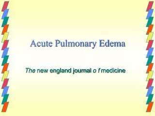
Acute Pulmonary Edema in Pregnancy • Cohort study – 62,917 consecutive pregnancies from 1989-1999, to describe the incidence, predisposing factors contributing to pulmonary edema in the pregnant patient. • Fifty-one women (0.08%) were diagnosed with acute pulmonary edema during ante partum - post partum period. • 24 patients (47%) antepartum • 7 patients (14%) intrapartum • 20 patients (39%) post partum • Most common causes: • Tocolytics (25.5%) most commonly MgSO4 and SC terbutaline • Cardiac disease (25.5%) • Fluid overload (21.5%) • Preeclampsia (18%)
Risk Factors • Preeclampsia/eclampsia • Tocolytic therapy • Sever infection • Cardiac disease • Iatrogenic fluid overload • Multiple gestation
EBM Discussion on Anesthetic Technique for Cesarean Section for Severe Preeclampsia • Randomized comparison of general and regional anesthesia for cesarean delivery in pregnancies complicated by severe preeclampsia (1995) • Eighty women who required C/S • Randomized - epidural/CSE/General • Intra-operative BP compared in all groups • No statistical or clinical difference in maternal or fetal outcome • Aside from logistical implications, general as well as regional were shown to be equally acceptable if steps are taken to ensure careful approach to either method.
EBM Discussion on Anesthetic Technique for Cesarean Section for Severe Preeclampsia • Prospective cohort study; Patients with severe preeclampsia experience less hypotension during spinal anesthesia for elective cesarean delivery than healthy parturients (2003). • Compared incidence and severity of spinal anesthesia assoaicated hypotension in severe preeclamptic (n=30) vs. healthy parturients (n=30). • Under spinal, mean BP decreased by 32-39% in severe preeclamptics and 33-60% in the healthy parturient. • Healthy patients were given more ephedrine than the preeclamptics for hypotension. Possible explained by increased sensitivity of pressor drugs in the preeclamptic. • Findings suggest that the incidenc of severity of spinal hypotension in preeclamptic patients with severe hypertension may be less than previously believed.
Discussion • Most likely cause of pulmonary edema is multifactorial. No specific etiology was assigned. • Unsuspected cardiac findings were common, and there was a high incidence of valvular disease. • Most pts had severe systolic dysfunction and LVH and not cardiac disease. • Underlying cardiac disease is most likely under-diagnosed and under-reported due to under-use of echocardiography.
References Journals • A. Sciscione et al. Acute Pulmonary Edema in Pregnancy. Obstetrics & Gynecology 2003;103:511-14. • D. Wallace et. al. Randomized Comparison of General and Regional Anesthesia for Cesarean Delivery in Pregnancies Complicated by Severe Preeclampsia. Obstetrics & Gynecology 1995;86:198-98. • A. Aya et al. Patients with Severe Preeclampsia Experience Less Hypotension During Spinal Anesthesia for Elective Cesarean Delivery than Healthy Parturients: A Prospective Cohort Comparison. Anesthesia & Analgesia 2003;97:867-72 Texts/Other • Baresh, Paul G. Clinical Anesthesia. • Stoelting, R. Anesthesia and Coexisting Disease • Upto Date – www.uptodate.com ; keyword – severe preeclampsia
Questions???? www.anaesthesia.co.inanaesthesia.co.in@gmail.com