RBCs Abnormal morphology
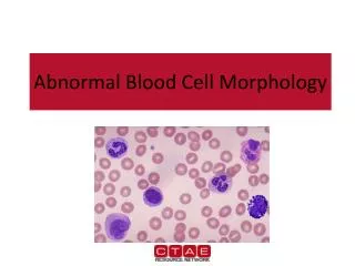
RBCs Abnormal morphology. Blood Cells. Abnormal erythrocyte morphology. Abnormal erythrocyte morphology is found in pathological states that may be : abnormalities in size (anisocytosis). abnormalities in shape (poikilocytosis).

RBCs Abnormal morphology
E N D
Presentation Transcript
RBCs Abnormal morphology Mohammed laqqan
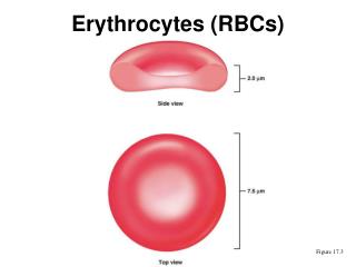
Blood Cells Mohammed laqqan
Abnormal erythrocyte morphology Abnormal erythrocyte morphology is found in pathological states that may be : • abnormalities in size (anisocytosis). • abnormalities in shape (poikilocytosis). • abnormalities in hemoglobin content or the presence of inclusion bodies in erythrocyte. Mohammed laqqan
I- Variation in red cells Distribution 1- Rouleaux Formation: • Morphology: Stacks of RBC's resembling a stack of coins. • Found in:- Hyperfibrinogenaemia- Hyperglobulinaemia Mohammed laqqan
I- Variation in red cells Distribution 2- Red cell-agglutination: • Morphology: Irregular clumps of red cells • Found in:- Cold agglutinins- Warm autoimmune hemolysis Mohammed laqqan
Terms Normochromic: A descriptive term applied to a red blood cell with a normal concentration of hemoglobin. Normocytic: A descriptive term applied to normal size of RBC. Hypochromic: A descriptive term applied to a red blood cell with a decreased concentration of hemoglobin. Macrocytic: A descriptive term applied to a larger than normal red blood cell. Mohammed laqqan
1-Microcytosis: Morphology: - Decrease in the red cell size. Red cells are smaller than ± 7µm in diameter. The nucleus of a small lymphocyte (± 8,µm) is a useful guide to the size of a red blood cell. Found in: - Iron deficiency anemia.- Thalassaemia.- Sideroblastic anemia.- Lead poisoning.- Anemia of chronic disease. II-Variation in erythrocyte size (anisocytosis) Mohammed laqqan
II-Variation in erythrocyte size (anisocytosis) Comment: Most erythrocytes presented in the picture are microcytes (compare with the small lymphocyte). The degree of hemoglobinization is sufficient. Normal platelets and single ovalocytes are present. Mohammed laqqan
II-Variation in erythrocyte size (anisocytosis) 2-Macrocytosis: • Morphology:Increase in the size of a red cell. Red cells are larger than 9µm in diameter. May be round or oval in shape, the diagnostic significance being different. • Found in:- Folate and B12 deficiencies (oval)- Ethanol (round)- Liver disease (round)- Reticulocytosis (round) Mohammed laqqan
III-Variation in hemoglobin content 1-Hypochromasia: • Morphology:Increase in the red cells' central pallor which occupies more than the normal third of the red cell diameter. • Found in:- Iron deficiency- Thalassaemia any of the conditions leading to Microcytosis Mohammed laqqan
III-Variation in hemoglobin content 2- Polychromasia: • Morphology:Red cells stain shades of blue-gray as a consequence of uptake of both eosin (by hemoglobin) and basic dyes (by residual ribosomal RNA). Often slightly larger than normal red cells and round in shape - round macrocytosis. • Found in:Any situation with reticulocytosis - for example bleeding, hemolysis or response to heamatinic factor replacement. Mohammed laqqan
IV- Variation of red cells shape (Poikilocytosis) 1- Spherocytosis: • Morphology:Red cells are more spherical. Lack the central area of pallor on a stained blood film. • Found in:- Hereditary spherocytosis.- Immune haemolytic anemia.- Zieve's syndrome (Look to the margin). - Microangiopathic haemolytic anemia. Mohammed laqqan
IV- Variation of red cells shape (Poikilocytosis) 2-Target Cells: • Morphology:Red cells have an area of increased staining which appears in the area of central pallor. • Found in:-Obstructive liver disease- Thalassaemia (not I.D.A)- Haemoglobinopathies (S and C)- Post splenectomy Mohammed laqqan
IV- Variation of red cells shape (Poikilocytosis) 3- Ovalocytes: Morphology:oval shape red blood cell Found in:- Thalassaemia major. - Hereditary ovalocytosis. - Sickle cell anemia Mohammed laqqan
IV- Variation of red cells shape (Poikilocytosis) 4- Elliptocytosis: • Morphology:The red cells are oval or elliptical in shape. Long axis is twice the short axis. • Found in: - Hereditary elliptocytosis- Megaloblastic anemia- Iron deficiency - Thalassaemia- Myelofibrosis Mohammed laqqan
IV- Variation of red cells shape (Poikilocytosis) 5- Tear Drop Cells: • Morphology:Red cells shaped like a tear drop or pear. • Found in: - Bone marrow fibrosis- Megaloblastic anemia- Iron deficiency- Thalassaemia Mohammed laqqan
IV- Variation of red cells shape (Poikilocytosis) 6- Blister cell (prekeratocyte): • Morphology: Have accentric hallow area. Resemble a women's handbag • Found in: Microangiopathic hemolytic anemia Mohammed laqqan
IV- Variation of red cells shape (Poikilocytosis) 7- Keratocytes (horn cell): • Morphology:Part of the cell fuses back leaving two or three horn-like projections. The keratocyte is a fragile cell and remains in circulation for only a few hours. • Found in:- Uraemia- Severe burns- EDTA artifact- Liver disease Mohammed laqqan
IV- Variation of red cells shape (Poikilocytosis) 8- Burr (crenation ) cell: Morphology: Red cell with uniformly spaced, pointed projections on their surface. Found in:- hemolytic anemia - Uremia. - Megaloblastic anemia Mohammed laqqan
IV- Variation of red cells shape (Poikilocytosis) 9- Acanthocytosis: • Morphology:are red blood cells with irregularly spaced projections, these projections very in width but usually contain a rounded end • Found in:- Liver disease - Post splenectomy- Anorexia nervosa and starvation Mohammed laqqan
IV- Variation of red cells shape (Poikilocytosis) 10- Schistocytosis: • Morphology: Fragmentation of the red cells. • Found in:- DIC - Micro angiopathic hemolytic anemia- Mechanical haemolytic anemia Mohammed laqqan
IV- Variation of red cells shape (Poikilocytosis) 11- Stomatocytosis: • Morphology:Red cells with a central linear slit or stoma. Seen as mouth-shaped form in peripheral smear. • Found in:- Alcohol excess- Alcoholic liver disease- Hereditary stomatocytosis- Hereditary spherocytosis Mohammed laqqan
IV- Variation of red cells shape (Poikilocytosis) 12- Sickle Cells: • Morphology: Sickle shaped red cells. • Found in: Hb-S disease Mohammed laqqan
IV- Variation of red cells shape (Poikilocytosis) 13- Nucleated red blood cells. These red blood cells are released from the bone marrow early into the blood stream, due to the need for oxygen. Normal red blood cells do not contain a nucleus on a peripheral smear. Mohammed laqqan
Envelope form cell • Found in • thalassemia • Sickle cell anemia Mohammed laqqan
V -Erythrocyte inclusion bodies 1- Howell-Jolly Bodies: • Morphology:Small round cytoplasmic red cell inclusion with same staining characteristics as nuclei • Found in:- Post splenectomy- Megaloblastic anemia Mohammed laqqan
V -Erythrocyte inclusion bodies 2- Siderotic Granules RBCs which contain no of hemoglobin iron granules. They appear as dense blue, irregular granules which are unevenly distributed in Wright stained RBCs. Pappenheimer bodies can be increased in hemolytic anemia, infections and post-splenectomy. Mohammed laqqan
V -Erythrocyte inclusion bodies 3- Basophilic stippling: • Morphology:Considerable numbers of small basophilic inclusions in red cells. • Found in:- Thalassaemia- Megaloblastic anemia- Hemolytic anemia - Liver disease- Heavy metal poisoning. Mohammed laqqan
V -Erythrocyte inclusion bodies 4- Heinz Bodies: • Represent denatured hemoglobin (methemoglobin - Fe+++) within a cell. With a supravital stain like crystal violet, Heinz bodies appear as round blue precipitates. • Presence of Heinz bodies indicates red cell injury and is usually associated with G6PD-deficiency. Mohammed laqqan
V -Erythrocyte inclusion bodies 5- Cabot Rings: • Reddish-blue threadlike rings in RBCs of severe anemia's. These are remnants of the nuclear membrane and appear as a ring or figure 8 pattern. • Very rare finding in patients with Megaloblastic anemia, severe anemia's, lead poisoning, and dyserythropoiesis. Mohammed laqqan
IV -Erythrocyte inclusion bodies 6- Parasites of Red Cell: • Are protozoan parasites which occur in many species of birds and are the cause of avian malaria. • Transmitted by mosquitoes, infection with Plasmodium spp can be a cause of hemolytic anemia Mohammed laqqan
RBCs Abnormal morphology Depiction of red blood cell morphologies that may appear on a peripheral smear, showing: (A) basophilic stippling, (B) Howell-Jolly bodies, (C) Cabot's ring bodies and (D) Heinz's bodies. Mohammed laqqan


![ERYTHROCYTES [RBCs]](https://cdn3.slideserve.com/5385917/erythrocytes-rbcs-dt.jpg)

![ERYTHROCYTES [RBCs]](https://cdn3.slideserve.com/6499114/erythrocytes-rbcs-dt.jpg)