CONJUNCTIVAL DISORDERS 5
330 likes | 513 Views
Learn about the definition, clinical manifestations, complications, diagnosis, and treatment of trachoma caused by Chlamydia trachomatis virus, impacting the conjunctiva and cornea.

CONJUNCTIVAL DISORDERS 5
E N D
Presentation Transcript
CONJUNCTIVAL DISORDERS5 BY Prof. Ahmad Mostafa
3. Chlamydial ConjunctivitisTrachoma • Definition • Aetiology • Clinical Manifestations • Sequelae (Complications) • Diagnosis • Treatment
Definition: Trachoma is a chronic infective disease which affects the conjunctiva and cornea. • It is characterized by formation of follicles, papillae, and pannus; and heals by cicatrization. • Aetiology: It is caused by an ‘atypical virus’ chlamydia trachomatis (TRIC agents), which produces intracytoplasmic basophilic inclusion bodies in the epithelial cells.
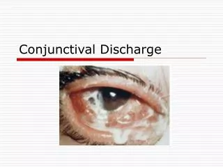
Clinical Manifestations • Incubation period: 6-12 days • Mode of onset: is insidious • Clinical manifestations include: (A) Conjunctival manifestations (B) Corneal manifestations
(A) Conjunctival manifestations of trachoma • Mac Callan’s classification (4 stages) First stage (T1) is characterized by the presence of the early (immature) follicles which are: 1. Minute up to 1 mm 2. Affect the upper palpebral conj. and fornix 3. Yellowish in color 4. Surrounded by dilated capillaries 5. Not raised, nor expressible 6. Scrabing of epithelium shows inclusion bodies
Second stage (T2 ) is further subdivided into: (a) T2a (Typical follicular trachoma) which is characterized by the presence of the mature follicles which are: 1. Large follicles 1-3 mm 2. Affect the upper palpebral conj. and fornix 3. Yellowish in color 4. Raised above the surface of the conj. 5. Expressible necrotic material 6. Scrabing of epithelium shows inclusion bodies
(b) T2b (Papillary trachoma): Papillae are fine finger-like processes. They are pink, highly vascular, and give the surface of the conj. a “velvety appearance”
Third stage (T3) (= Cicatrising trachoma) Fibrosis starts to appear in the tarsal conj. and manifested by: • Arlt’s line: a band of fibrous tissue occupying the sulcus subtarsalis (most vascular part of the conj). It is pathognomonic of trachoma. • Post-trachomatous degenerations (PTD) result from hyaline degeneration in a closed crypt (between papillae). They appear yellow in color. • Post-trachomatous concretions (PTC) are white in color and result from calcification of PTD. N.B. With the onset of fibrosis, trichiasis, entropion …etc occur hence this stage is called “ stage of complications”
Fourth stage (T4) (= Healed trachoma) The patient is cured and not infective. Scrabings from the conjunctiva show no inclusion bodies.
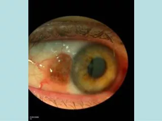
(B) Corneal manifestations of trachoma • Corneal follicles • Trachomatous pannus • Trachomatous ulcers
1. Corneal follicles • They form in the upper part of the cornea commonly in association with pannus. • They are small, greyish in color. • Are present between the epithelium and Bowman’s membrane. • They are called ( Herbert’s rosettes ). • When heal by fibrosis leave depressed pits in the upper cornea (Herbert’s pits).
2. Trachomatous pannus • Definition • Course • Types • Fate • Differential Diagnosis
Definition: Pannus means vascularization + cellular infiltration of the upper part of the cornea.
Course:is as follows: • Progressive pannus: Cellular infiltration occurs in the upper part of the cornea between epithelium & BM, followed by superficial vascularization i.e. cellular infiltration precedes the vascularization. 2. Regressive pannus: cellular infiltration regresses but the vessels never regress, thus, the vessels are seen extending a short distance infront of the cellular infiltration. 3. Healed pannus (= Dry pannus = pannus siccus) A superficial scar is formed in the upper part of the cornea. Herbert’s pits may be seen with pannus giving it a serrated appearance.
Types: 1. Pannus tenius = Thin pannus 2. Pannus carnosus = Fleshy pannus (cellular infiltration is excessive) 3. Pannus vasculosus = Vascular pannus (vascularization is excessive) 4. Pannus annulosus = Annular pannus (affects the cornea all around) 5. Pannus Siccus = healed pannus
Fate of pannus: • Complete resolution if B.M. is not destroyed • Opacity in the upper cornea if B.M. is destroyed pannus siccus
3. Trachomatous ulcers • Trachomatous ulcers are of 3 types: • Ulcers related to pannus either at the edge or on the surface of pannus. It is linear and horizontal (typical trach. ulcer). • Ulcers unrelated to pannus may be central or marginal. • Ulcers produced mechanically by trichiasis, PTD …etc
Sequelae (Complications)Of Trachoma • Lid complications • Conjunctival complications • Corneal complications • Lacrimal complications
Lid complications 1. Rubbing lashes or trichiassis: • From fibrosis around the hair follicles • From growth of new abnormal follicles due to chronic hyperemia • Associated with entropion caused by tarsal conj. fibrosis. 2. Cicatricial entropion 3. Chronic blepharitis 4. Recurrent chalazia due to fibrosis of ducts of Meibomian glands 5. Ptosis: - Early due to: a. Infiltration of Muller’s muscle b. Mechanical (thickened conj.) - Late: from fibrosis of Muller’s muscle
Conjunctival complications • Posterior symblepharon due to fibrosis obliteration of the fornices • Xerosis: results from: a. Atrophy of goblet cells of the conj. b. Stenosis ( obstruction) of lacrimal ductules by fibrosis. • Hyaline & amyloid degeneration of the conj. • Epithelial plaque formation
Corneal complications • Corneal ulcers: 3 types mentioned before • Xerosis • Keratectasia ex pano • Epithelial plaque • Diffuse opacities
Lacrimal complications • Punctal stenosis • Canalicular stenosis • Fibrosis of the fornix occlusion of lacrimal ductules • Chronic dacryocysytis (from obstruction of NLD) • Chronic dacryoadenitis
Diagnosis of trachoma • Clinical signs: pannus, Arlt’s line, Herbert’s pits. • Epithelial scrapings stained with Giemsa stain showing the characteristic Halberstaedter-Prowazek bodies (intracytoplasmic basophilic inclusion bodies) • Immunofluorescent staining of conj. smears • Cultivation of TRIC agents on yolk sac of chick embryo.
Treatment of Trachoma • Medical ttt: • Systemic a. Tetracycline 250 mg tid daily for 1 month b. Erythromycin for children below 8 (tetracycline affects teeth & bone growth) • Topical a. Sulfacetamide 20 or 30% 5 times daily b. Terramycin eye ointment at bed time
Surgical ttt: • Mature follicles expression • PTD picking • Hyaline and amyloid degeneration with very thickened conj. combine excision of tarsus and conj.
Differential Diagnosis of A Red Eye • Acute Conjunctivitis • Acute Keratitis & corneal ulcer • Acute iridocyclitis • Acute congestive glaucoma • Subconjunctival hemorrhage