Download
1 / 66
660 likes | 956 Views
CONJUNCTIVAL DEGENARATIONS AND DRY EYES. Dr. Faizur Rahman Professor of Ophthalmology Peshawar Medical College. CONJUNCTIVAL DEGENERATIONS. Pinguecula. Pterygium. Xerosis. Concretions. Retention cysts. PINGUECULA.

E N D
CONJUNCTIVAL DEGENARATIONS AND DRY EYES Dr. Faizur Rahman Professor of Ophthalmology Peshawar Medical College.
CONJUNCTIVAL DEGENERATIONS • Pinguecula. • Pterygium. • Xerosis. • Concretions. • Retention cysts.
PINGUECULA • A pinguecula is a benign degenerative tumor, appear as localized elevated area in the interpalpebral fissure on the limbal conjunctiva. • Nasal & bilateral. • Yellow, gray, white, or colorless. • Chronic exposure to the sun. • There is no predilection for sex or race.
PATHOPHYSIOLOGY • Exposure (Toxic vapors, salt water spray, sun). • Insufficient moisture and lubrication (tears). • Elastotic degeneration and deposition of abnormal elastic fibers in the conjunctival substantia propria. • Heredity. • Heat, dryness, wind, dust, smoke, and other irritants.
CLINICAL FEATURES • In most cases, pingueculae are an ancillary finding. • Cosmetic defect. • Corneal punctate epitheliopathy and dellen. • Pingueculitis. • Conjunctiva may become irritated.
MANAGEMENT • Mostly it is asymtomatic, so no intervention. • Prevention of exposure. • Lubricants. • Steroids. • Surgical resection.
PTERYGIUM • Pterygia are triangular, fibrovascular connective tissue overgrowths of bulbar conjunctiva onto the cornea. • They are horizontally located in the interpalpebral fissure. • Warm, dry climates, or chronical exposure to the sun or smoky/dusty environments. • Pingueculae may precede pterygia.
PATHOPHYSIOLOGY • Drying of the interpalpebral tear film is an important factor. • This exposes the peripheral cornea to destructive effects of the UV light, and the tissue damage thus sustained stimulates the advance of limbal vessels into the cornea. • Drying of interpalpebral film occurs most readily in the medial third of the I/P fissure.
PATHOPHYSIOLOGY • Ultraviolet light exposure (both UV-A and UV-B). • Corneal stromal edema. • Invasion by blood vessels and fibroblast. • Organization of this fibrovascular tissue. • Allergens, noxious chemicals and irritants (e.g., wind, dirt, dust, and air pollution). • Heredity may also be a factor.
HISTOLOGY • Degeneration of the conjunctival stroma with its replacement by thickened, tortuous elastotic fibers. • Actinically activated fibroblasts invade and fragment Bowman's layer. • Except at its apex, the pterygium is covered by conjunctival epithelium. • Histologically, pterygium development resembles actinic degeneration of the skin.
CLINICAL FEATURES • Cosmetic concerns and surface irritation are the most common complaints. • In most cases pterygia are asymptomatic. • Vascularized pterygium may become red and inflamed. • Irregular ocular surface can interfere with the stability of the precorneal tear film.
CLINICAL FEATURES • Pterygium may effect vision. • Stocker's line. • Persistent foreign body sensation. • Diplopia. • A pterygium may progress slowly toward axial cornea or may become quiescent.
DIFFERETIAL DIAGNOSIS • Pinguecula. • Pseudopterygium. • Conjunctival intraepithelial neoplasia (CIN). • Pyogenic granuloma.
MANAGEMENT • Avoidance of the causative factors. • Topical decongestant / antihistamine combinations and/or mild topical steroids. • Surgery
MANAGEMENT…Cont. Surgical excision of pterygia is indicated only for: Unacceptable cosmoses. Significant encroachment of the visual axis or there is significant astigmatism. A persistent foreign body sensation in the eye. Constant or recurrent inflammation and irritation. Restriction of extraocular muscle movement.
SURGICAL REMOVAL • Surgery is the only way to remove a pinguecula or pterygium. • The recurrence rate is often as high as 50 to 60 percent. • Procedure and outcome.
ARGON LASER PHOTOCAGULATION • Laser treatment early on and for recurrence has been helpful. • Has the advantage of regressing repeated growths for long periods. • The treatment is done in one or two sessions and it is split in two phases: Phase 1: • Direct photocoagulation of the largest vessels. • 50-100 micron spot.
ARGON LASER PHOTOCAGULATION Phase 2: • The ray is defocused into the body of the growth by using larger diameters (200-300 micron) and less power (150-300 mW) in order to cause a contraction of the fibrous/elastic subconjunctival tissue. • Stable regression in 90% cases has been reported two years after the treatment, and in the case of recurrences, the lesion had a very slow and benign evolution.
RECURRENT PTERYGIUM • Pterygia often persist after surgical removal; These "recurrent pterygia" probably have no relationship to ultraviolet radiation, but rather may be likened to keloid development in the skin. The rate of recurrence is significantly high 50 - 60 percent when a bare sclera excision is performed. • Treatment with autologous conjunctival transplantation has been shown to decrease the incidence of recurrence to about 5 percent.
RECURRENT PTERYGIUM • This rate is usually reduced if surgery is followed by beta-irradiation with strontium 90. But many complications. • Adjunctive treatment with mitomycin drops or Thiotepa. • In cases that involve significant corneal scarring, lamellar or penetrating keratoplasty may be indicated. • Follow up for pterygia or recurrence is at least once or twice yearly, and include a manifest refraction, corneal topography, slit lamp evaluation with measurement of the pterygium, and photodocumentation if possible.
XEROSIS • Abnormal lid movement, tear hyposecretion (keratoconjunctivitis sicca), or mucus deficiency. Malnutrition. • Epidermalization with keratin formation. • Xerophthalmia, the result of prolonged deficiency of Vitamin A. • Loss of the mucus-secreting conjunctival goblet cells • Squamous metaplasia of conjunctival epithelial cells. • Conjunctival xerosis is typically bulbar in distribution. • Bitot's spots. • Conjunctival xerosis and Bitot's spots can be reversed.
CONCRETIONS/ LITHIASIS • Degenerations of conj. epithelium in elderly or prolonged conjunctivitis or meibomian gland disease may cause yellowish to white concretions in the epithelium. • The deposits may be seen as linear streaks in the palpebral conjunctiva or as minute spheres in the inferior fornix. • FB sensation.
RETENTION CYST • Lymphatic channels of the conjunctiva may become dilated and cause serous conjunctival cysts filled with clear fluid. • Mostly asymptomatic and if indicated can be punctured with a needle.
DRY EYES • Dry eye syndrome (DES) is a common disorder • Quantitative or qualitative deficiency in the tear film.
DRY EYES • 3 Layers of tear film: • Lipid layer --- 0.11 microns. Meibomian glands. • Aqueous layer --- 7.0 microns. Lacrimal glands. • Mucin layer ---0.02-0.5 microns. Goblet cells. • Defective or deficient tear film will result in a dry eye.
CAUSES • Aqueous tear layer ( KCS): 1. CONGENITAL: • Aplasia or hypoplasia of lacrimal gland. • Riley-day syndrome (Dysautonomia). • Anhidrotic ectodermal dysplasia. • Aplasia of lacrimal nerve nucleus. • Multiple endocrine neoplasia.
CAUSES (contd.) 2. ACQUIRED: • Senile or idiopathic atrophy of lacrimal gland. • Postsurgical.(Blepharoplasty,Dacryoadenectomy) • Traumatic, inflammatory or neoplastic lesions of lacrimal gland. • Neuro-paralytic lesions: Facial nerve, Geniculate ganglion, Spheno-palatine ganglion, Greater Superficial petrosal nerves, Trigeminal nerve and Gasserian ganglion. • Nutritional and debilitating disorders : Typhus, cholera, starvation, ascorbic acid and vit. B12 deficiency.
CAUSES (contd.) • Systemic Diseases: • Connective tissue disorders (R.A, SLE, Periarteritis nodosa, Scleroderma). • Endocrine disorders (Hashimoto‘s disease, Menopause). • Renal diseases (Renal tubular acidosis, Diabetes insipidus). • Blood disorders (Hemolytic anemia, Hypergammaglobinemia, Felty‘s syndrome, Malignant lymphoma, Lymphoid leukemia, Lymphosarcoma, Chronic hepatitis, Primary biliary cirrhosis). • Skin and muco-cutaneous disorders (Sclerodera, erythema multiforme, Exfoliative dermatitis, Cicatricial pemphigoid). • Miscellaneous (Sarcoidosis, Amyloidosis, Lipodystrophy)
CAUSES (contd.) • Mucin tear layer: • Vit. A deficiency. • Trachoma. • Diphtheric kerato-conjunctivitis. • Chemical, thermal and radiation injuries of conjunctiva. • Topical medications—Echothiophate iodide, Sulphonamides etc
Causes (contd.) • Lipid tear layer: • Chronic conjunctivitis. • Acne rosacea. Other and newer causes: *After cataract extraction *After PRK *Contact lens wear
Associated with: • Connective tissue diseases • Steven Johnson syndrome • Vit. A deficiency • AIDS • Hepatitis C • Polycystic ovarian syndrome • Post radiation (damage to salivary gland)
VITAMIN A DEFICIENCY • Nactylopia is often the presenting symptom. • Decreased mucus production by goblet cells. • Xerosis—Dryness of conjunctiva and cornea. • Bitot‘s spot---Metaplastic keratinization of areas of conjunctiva. • Corneal ulcers and scars. • Kerato-malacia, corneal necrosis.
VITAMIN A DEFICIENCY • Bitot‘s spot is a superficial, foamy, triangular area in the conjunctiva, in the interpalpebral aperture. It consists of keratinized epithelium, inflammatory cells, debris and Coryne - bacterium Xerosis. • Acute vit. A deficiency (keratomalacia) is a medical emergency with an untreated mortality rate of 50%.
SJOGREN SYNDROME • PRIMARY: Aqueous tear deficiency associated with dry mouth (xerostomia). • Serology for • Rh factor • Antinuclear antibody • Salivary gland Biopsy.
SJOGREN SYNDROME • SECONDARY: Aqueous tear deficiency associated with definite Connective tissue disease. • Multisystem autoimmune disease, most commonly associated with Rh.arthritis. • There is an autoimmune infiltration of lacrimal and salivary glands by lymphocytes. • Lacrimal gland biopsy.
MIKULICZ‘S SYNDROME • Enlargement of lacrimal / salivary glands or both, owing to systemic diseases, such as Leukemia, Lymphoma or Sarcoidosis.
DRUGS • Many systemic drugs can decrease aqueous tear production: • Antihistamines, • Hypnotics, • Phenothiazines, • Psychotropics, • Halothane, • Antimuscarinics (atropine), • Beta blockers (timolol), • Hexamethonium, • Nitrous Oxide etc.
CLINICAL FEATURES OF DRY EYES • SYMPTOMS: • Grittiness, Itching, Burning sensation, Foreign body sensation and photophobia • Redness of the eyes. • Reflex lacrimation. • Blurred vision. • Stringy mucus secretion. • Severe pain ( Filamentary keratopathy ).
CLINICAL FEATURES OF DRY EYES • Worsening Factors: • Prolonged use of eyes e.g; prolonged reading, watching TV, excessive computer use. • Symptoms worsen in the morning and towards the end of the day. • Temperate climate, during winter. • Lower levels of humidity (Indoor heating systems ), smoky and dry environment like kitchen, busy street and outdoor work. • Air-conditioned atmosphere
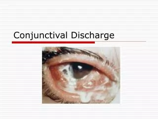
CLINICAL FEATURES OF DRY EYES • SIGNS: • Decreased tear meniscus and irregular edges or scalloped appearance along the lid margin. Normal height --- 1mm. Concave tear meniscus. • Viscous and stringy mucous due to debris in the tear film. • Increased debris and foam in tear film. • Xerosis ( dry conjunctiva ) and Bitot‘s spots. • Hyperemia and Papillary conjunctivitis
CLINICAL FEATURES OF DRY EYES • Irregular corneal surface ( fine, granular, coarse or confluent epithelial keratopathy ). • Severe cases--- Corneal ulcer and bacterial colonization causing suppurative keratitis --- Perforation. • Secondary infection is also aided by deficiency of lysozyme and other antibacterial agents. • Inadequate or insufficient blinking. • Filaments and mucous plaques. Filaments are strands of epithelial cells attached to the surface of cornea. Painful.